Impaction of an intrathoracic kidney acted as a shield against herniation of the abdominal viscera in a case of right congenital diaphragmatic hernia
- Affiliations
-
- 1Department of Obstetrics and Gynecology, University of Ulsan College of Medicine, Asan Medical Center, Seoul, Korea. jyshim@amc.seoul.kr
- KMID: 2152646
- DOI: http://doi.org/10.5468/ogs.2016.59.1.58
Abstract
- We describe a case of an intrathoracic kidney combined with right congenital diaphragmatic hernia (CDH) that was diagnosed at 32 weeks of gestation. Although it has been well established that a right CDH shows a poorer outcome than a left CDH, our present case showed a good outcome because there was no herniation of other abdominal viscera, except for the right kidney. Our findings in this case indicate that impaction of the intrathoracic kidney may act as a 'shield' against further herniation of other abdominal viscera into the thoracic cavity.
Figure
-
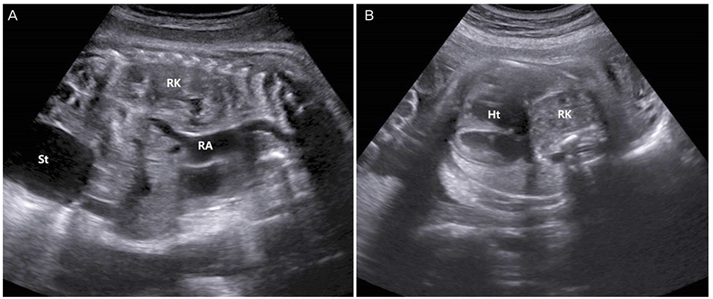
Fig. 1 Ultrasonographic findings for the study patient at 32 weeks of gestation. (A) Parasagittal view of the fetus showing a mixed echogenic mass (RK) behind the right atrium (RA) of the fetal heart. The fluid-filled stomach (St) was in a normal position. (B) Subcostal 4-chamber view of the fetal heart (Ht) showing a mesocardia, with rotation of the heart and its apex located in the mid-thorax. A mixed echogenic right kidney was located behind the right side of the fetal heart.
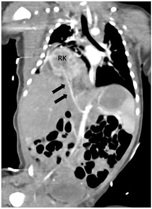
Fig. 2 By postnatal computed tomography, through the right congenital diaphragmatic defect, a posterolateral herniated right kidney (RK) was observed in the neonatal chest and a right renal artery (black arrows) was stretched to feed the intrathoracic kidney.
Reference
-
1. Beaumier CK, Beres AL, Puligandla PS, Skarsgard ED. Canadian Pediatric Surgery Network. Clinical characteristics and outcomes of patients with right congenital diaphragmatic hernia: a population-based study. J Pediatr Surg. 2015; 50:731–733.2. DeKoninck P, Gomez O, Sandaite I, Richter J, Nawapun K, Eerdekens A, et al. Right-sided congenital diaphragmatic hernia in a decade of fetal surgery. BJOG. 2015; 122:940–946.3. Fisher JC, Jefferson RA, Arkovitz MS, Stolar CJ. Redefining outcomes in right congenital diaphragmatic hernia. J Pediatr Surg. 2008; 43:373–379.4. Hedrick HL, Crombleholme TM, Flake AW, Nance ML, von Allmen D, Howell LJ, et al. Right congenital diaphragmatic hernia: prenatal assessment and outcome. J Pediatr Surg. 2004; 39:319–323.5. Skari H, Bjornland K, Haugen G, Egeland T, Emblem R. Congenital diaphragmatic hernia: a meta-analysis of mortality factors. J Pediatr Surg. 2000; 35:1187–1197.6. Donat SM, Donat PE. Intrathoracic kidney: a case report with a review of the world literature. J Urol. 1988; 140:131–133.7. Park BS, Cho HJ, Ji YI, Jeong CH, Chun SW, Jeon GH, et al. A case of prenatal diagnosis of congenital thoracic kidney in the third trimester with congenital diaphragmatic hernia. Korean J Obstet Gynecol. 2012; 55:978–981.8. Athanasiadis AP, Zafrakas M, Arnaoutoglou C, Karavida A, Papasozomenou P, Tarlatzis BC. Prenatal diagnosis of thoracic kidney in the 2nd trimester with delayed manifestation of associated diaphragmatic hernia. J Clin Ultrasound. 2011; 39:221–224.9. Panda B, Rosenberg V, Cornfeld D, Stiller R. Prenatal diagnosis of ectopic intrathoracic kidney in a fetus with a left diaphragmatic hernia. J Clin Ultrasound. 2009; 37:47–49.10. Masturzo B, Kalache KD, Cockell A, Pierro A, Rodeck CH. Prenatal diagnosis of an ectopic intrathoracic kidney in right-sided congenital diaphragmatic hernia using color Doppler ultrasonography. Ultrasound Obstet Gynecol. 2001; 18:173–174.11. Hidaka N, Fujita Y, Satoh Y, Fukushima K, Wake N. Sonographic appearance of intrathoracic kidney in a fetus with left diaphragmatic hernia. J Clin Ultrasound. 2012; 40:600–602.12. Sherer DM, Eglinton GS, Goncalves LF, Lewis KM, Queenan JT. Prenatal color and pulsed Doppler sonographic documentation of intrathoracic umbilical vein and ductus venosus, confirming extensive hepatic herniation in left congenital diaphragmatic hernia. Am J Perinatol. 1996; 13:159–162.13. Harrison MR, Keller RL, Hawgood SB, Kitterman JA, Sandberg PL, Farmer DL, et al. A randomized trial of fetal endoscopic tracheal occlusion for severe fetal congenital diaphragmatic hernia. N Engl J Med. 2003; 349:1916–1924.14. Wilson JM, Lund DP, Lillehei CW, Vacanti JP. Congenital diaphragmatic hernia: a tale of two cities: the Boston experience. J Pediatr Surg. 1997; 32:401–405.15. Harrison MR, Adzick NS, Estes JM, Howell LJ. A prospective study of the outcome for fetuses with diaphragmatic hernia. JAMA. 1994; 271:382–384.
- Full Text Links
-
- Actions
-
Cited
- CITED
-
- Close
- Share
-
- Similar articles
-
- A case of prenatal diagnosis of congenital thoracic kidney in the third trimester with congenital diaphragmatic hernia
- A Case of Congenital Diaphragmatic Hernia that Occurred Following In Vitro Fertilization and Embryo Transfer
- Right-side Bochdalek Hernia with Unusual Kidney Herniation in an Old Patient
- Acute Pancreatitis Associated with Diaphragmatic Hernia in an Adult
- Unilateral Congenital Diaphragmatic Eventration Mimicking Congenital Diaphragmatic Hernia
