Korean J Obstet Gynecol.
2011 Mar;54(3):155-158. 10.5468/KJOG.2011.54.3.155.
A case of amniotic band syndrome associated with anencephaly demonstrated using three-dimensional ultrasound at early pregnancy
- Affiliations
-
- 1Department of Obstetrics and Gynecology, Haeundae Paik Hospital, Inje University College of Medicine, Busan, Korea. lory1202@naver.com
- 2Department of Pathology, Haeundae Paik Hospital, Inje University College of Medicine, Busan, Korea.
- KMID: 2274031
- DOI: http://doi.org/10.5468/KJOG.2011.54.3.155
Abstract
- Amniotic band syndrome (ABS) is entrapment of fetal parts by disrupted amnion and formation of fibrous amniotic band. As results, it causes a variety of fetal malformation involving the limbs, the craniofacial region and trunk. A visualization of free floating amniotic band is appropriate diagnostic method for ABS. Three dimensional (3D) and four dimensional (4D) ultrasounds are useful to diagnosis ABS. However prenatal diagnosis is difficult especially when they do not circulate a limb or cause single abnormality. In the cases of anencephaly, a differentiation between ABS and primary neural tube defect is important because the recurrence rate is significantly different. We reported a case that a fetal anencephaly with hydrops was diagnosed using prenatal 3D and 4D ultrasound then confirmed as amniotic band syndrome by autopsy at 12 weeks of gestation. After autopsy, we demonstrated amniotic band through 3D ultrasound review.
MeSH Terms
Figure
-
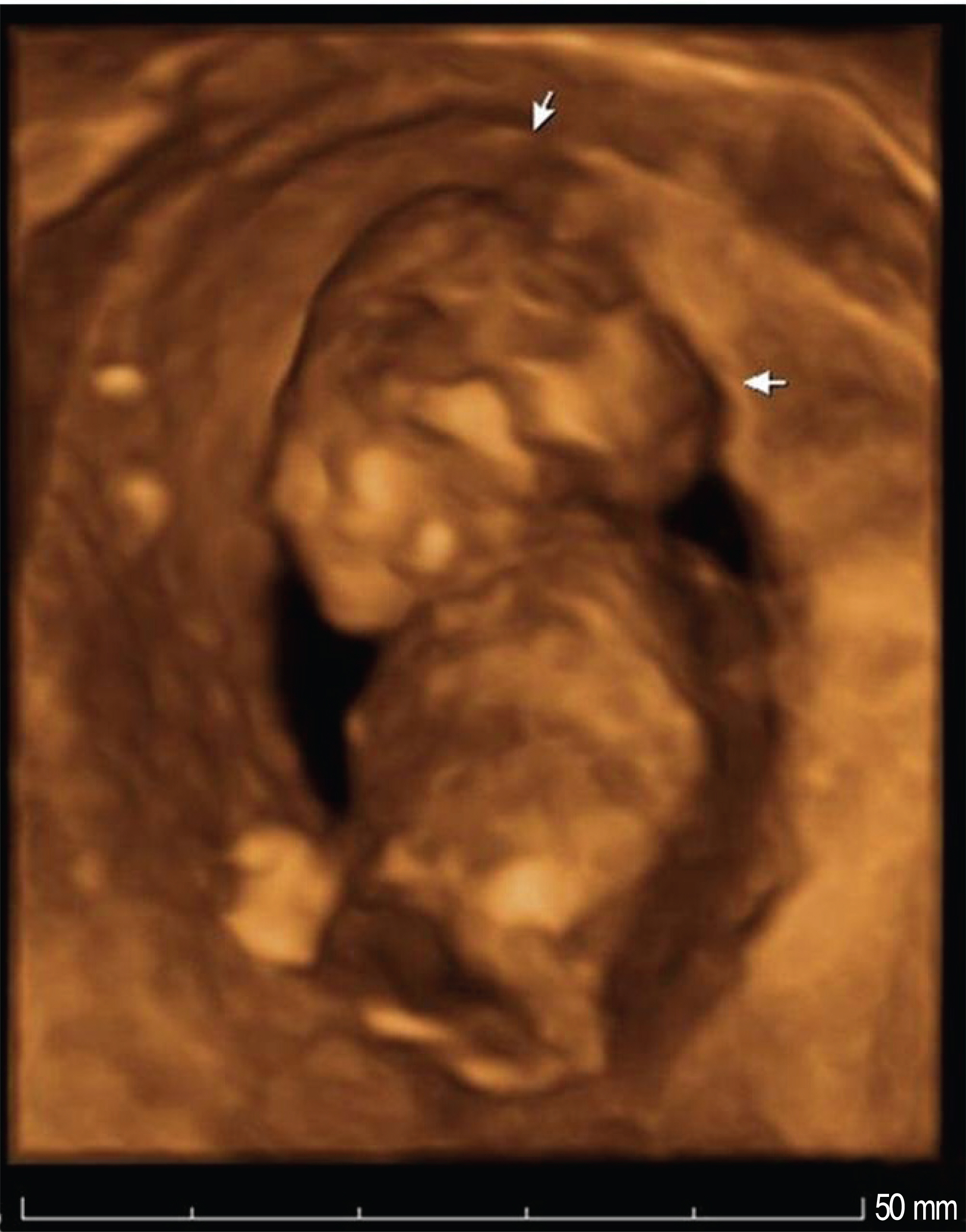
Fig. 1. 3D ultrasound of fetus with acrania. Calvarium is absent and remaining brain tissue shows abnormal contour (white arrow).

Fig. 2. (A) Autopsy finding of fetus and placenta. Amniotic membrane is attached to the fetal head. Fetal head and trunk are tilted to the left side. (B) Autopsy finding of fetus and placenta shows that amniotic membrane is attached to the segmented left thumb. (C) The posterior view of fetus with amniotic band.
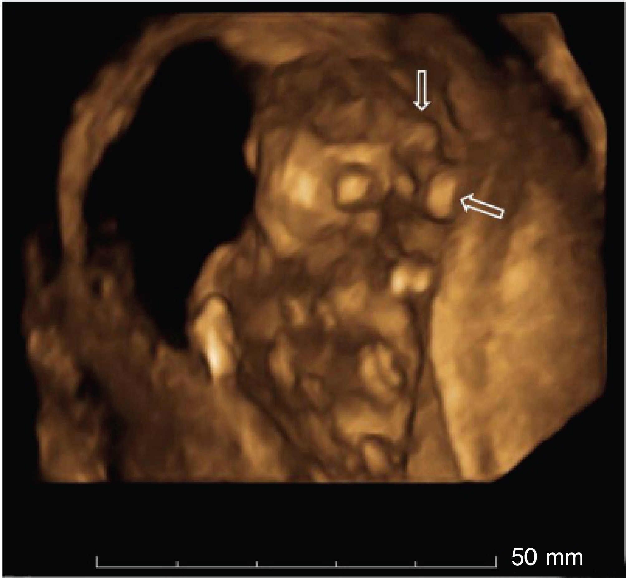
Fig. 3. Amniotic band between fetal head and placenta is demonstrated with review of three-dimensional surface rendering mode (white open arrow).
Reference
-
1. Garza A, Cordero JF, Mulinare J. Epidemiology of the early amnion rupture spectrum of defects. Am J Dis Child. 1988; 142:541–4.
Article2. Muraskas JK, McDonnell JF, Chudik RJ, Salyer KE, Glynn L. Amniotic band syndrome with significant orofacial clefts and disruptions and distortions of craniofacial structures. J Pediatr Surg. 2003; 38:635–8.
Article3. Rohrbach M, Chitayat D, Drake J, Velsher L, Sirkin WL, Blaser S. Prenatal diagnosis of fetal exencephaly associated with amniotic band sequence at 17 weeks of gestation by fetal magnetic resonance imaging. Fetal Diagn Ther. 2007; 22:112–5.
Article4. Laberge LC, Ruszkowski A, Morin F. Amniotic band attachment to a fetal limb: demonstration with realtime sonography. Ann Plast Surg. 1995; 35:316–9.5. Chen CP, Chang TY, Lin YH, Wang W. Prenatal sonographic diagnosis of acrania associated with amniotic bands. J Clin Ultrasound. 2004; 32:256–60.
Article6. Paladini D, Foglia S, Sglavo G, Martinelli P. Congenital constriction band of the upper arm: the role of three-dimensional ultrasound in diagnosis, counseling and multidisciplinary consultation. Ultrasound Obstet Gynecol. 2004; 23:520–2.
Article7. Inubashiri E, Hanaoka U, Kanenishi K, Yamashiro C, Tanaka H, Yanagihara T, et al. 3D and 4D sonographic imaging of amniotic band syndrome in early pregnancy. J Clin Ultrasound. 2008; 36:573–5.
Article8. Chen HE, Chen CP, Hsu CY, Wang W. Typical body wall defect associated with craniofacial anomalies and amniotic bands diagnosed in early pregnancy. Taiwan J Obstet Gynecol. 2007; 46:286–7.
Article9. Pedersen TK, Thomsen SG. Spontaneous resolution of amniotic bands. Ultrasound Obstet Gynecol. 2001; 18:673–4.
Article10. Soldado F, Aguirre M, Peiro JL, Carreras E, Arevalo S, Fontecha CG, et al. Fetoscopic release of extremity amniotic bands with risk of amputation. J Pediatr Orthop. 2009; 29:290–3.
Article11. Husler MR, Wilson RD, Horii SC, Bebbington MW, Adzick NS, Johnson MP. When is fetoscopic release of amniotic bands indicated? Review of outcome of cases treated in utero and selection criteria for fetal surgery. Prenat Diagn. 2009; 29:457–63.12. Peiro JL, Carreras E, Soldado F, Sanchez-Duran MA, Aguirre M, Barber I, et al. Fetoscopic release of umbilical cord amniotic band in a human fetus. Ultrasound Obstet Gynecol. 2009; 33:232–4.
Article
- Full Text Links
-
- Actions
-
Cited
- CITED
-
- Close
- Share
-
- Similar articles
-
- The Amniotic Band Syndrome as a Cause of Anencephaly and Nuchal Skin Defect
- Prenatal Diagnosis of Exencephaly as a Consequence of an Amniotic Band Syndrome
- A case of amniotic band syndrome with anencephaly
- A Case of Amniotic Band Syndrome as a Cause of Anencephaly
- Antenatal Sonographic Diagnosis of the Amniotic Band Syndrome
