Korean Circ J.
2008 Oct;38(10):570-571. 10.4070/kcj.2008.38.10.570.
Coronary Artery-Right Ventricular Fistulae After an Acute ST Segment Elevation Myocardial Infarction
- Affiliations
-
- 1Division of Cardiology, Department of Internal Medicine, Pusan National University Hospital, Busan, Korea. glaraone@hanmail.net
- KMID: 2225724
- DOI: http://doi.org/10.4070/kcj.2008.38.10.570
Abstract
- No abstract available.
MeSH Terms
Figure
-
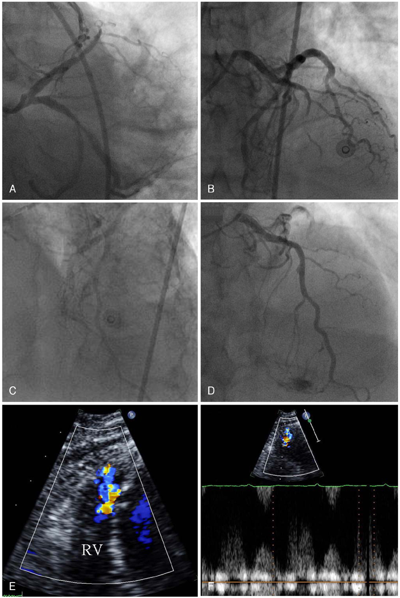
Fig. 1 The coronary angiography revealed a total occlusion of the middle left anterior descending coronary artery (LAD) and a fistulae originating from the proximal LAD to the main pulmonary artery (A and B). No abnormalities were observed in the distal LAD after the PCI (C). The follow up coronary angiography showed a fistulae from the septal branches of the distal LAD to the right ventricle (RV) and mild aneurysmal changes at the stent in the middle LAD after 9 months (D). The transthoracic echocardiography revealed a focal diastolic flow within the RV chamber (E and F). PCI: percutaneous coronary intervention.
Reference
-
1. Yu R, Sharma B, Franciosa JA. Acquired coronary artery fistula to the left ventricle after acute myocardial infarction. Am J Cardiol. 1986. 58:557–558.2. Cheong ER, Park HS, Yang DH, et al. Clinical characteristics of the patients with myocardial rupture after acute myocardial infarction. Korean Circ J. 2002. 32:467–472.3. Schanzenbächer P, Bauersachs J. Acquired right coronary artery fistula draining to the right ventricle: angiographic documentation of first appearance following reperfusion after acute myocardial infarction, with subsequent spontaneous closure. Heart. 2003. 98:e22.4. Ryan C, Gertz EW. Fistual frome coronary arteries to left ventricle after myocardial infarction. Br Heart J. 1977. 39:1147–1149.
- Full Text Links
-
- Actions
-
Cited
- CITED
-
- Close
- Share
-
- Similar articles
-
- Precordial ST-Segment Elevation in Acute Right Ventricular Myocardial Infarction
- Acute Myocardial Infarction by Right Coronary Artery Occlusion Presenting as Precordial ST Elevation on Electrocardiography
- Differences in Clinical Outcomes Between Patients With ST-Elevation Versus Non-ST-Elevation Acute Myocardial Infarction in Korea
- ST segment
- The Eletrocardiographic Analysis of Acute Myocardial Infarction and Non-infarction Syndrome In the Patients with ST Segment Elevation and Chest Pain
