J Korean Ophthalmol Soc.
2013 Apr;54(4):552-556. 10.3341/jkos.2013.54.4.552.
Comparison of Bacterial Culture Rate and Bacterial Floral Distribution in Anophthalmic Patients with Prosthetic Eye, Between Patients with Symptom and Without Symptom
- Affiliations
-
- 1Department of Ophthalmology, Chonbuk National University Medical School, Jeonju, Korea. ahnmin@jbnu.ac.kr
- KMID: 2216933
- DOI: http://doi.org/10.3341/jkos.2013.54.4.552
Abstract
- PURPOSE
To evaluate the distribution of conjunctival bacterial flora in anophthalmic socket patients with a prosthetic eye, and compare the bacterial positive culture rates between patients with subjective symptoms such as eye wax or irritation and patients without symptoms.
METHODS
Twenty-six anophthalmic socket patients with a prosthetic eye who visited our clinic between December 2009 and May 2011 were retrospectively analyzed. The patients were asked about their symptoms, followed by a conjunctiva examination. Specimens were obtained from the inferior conjunctival cul- de- sac with a sterile cotton-tipped applicator. The collected specimens were cultured.
RESULTS
The results indicated that the overall positive culture rate in the anophthalmic conjunctival socket was 69.2%, and the predominant organism was S. epidermidis (38.5%). Potential pathogenic bacteria were found in 4 eyes with a 15% positive culture rate. The incidence of bacteria was significantly higher (85.4%) in patient samples with subjective symptoms compared to patients without symptoms (50%). The bacterial positive culture rate of the potential pathogen bacteria in the group with symptoms was higher at 21%, but was not statistically significant.
CONCLUSIONS
Performing a pathogen culture test is necessary for prosthetic eye patients who complain of their symptoms. Additionally, the proper antibacterial treatment should be performed according to the antibiotics sensitivity of cultured bacteria.
Keyword
MeSH Terms
Figure
-

Figure 1. Correlation between bacterial positive culture rate and causes of enucleation.
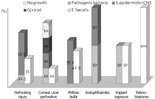
Figure 2. Correlation between bacterial species and causes of enucleation.
Reference
-
References
1. Fahmy JA, Moller S, Bentzon MW. Bacterial flora of the normal conjunctiva. Ⅰ. Topographical distribution. Acta Ophthalmol. 1974; 52:786–800.
Article2. Srinivasan BD, Jakobiec FA, Iwamoto T, DeVoe AG. Giant papillary conjunctivitis with ocular prostheses. Arch Ophthalmol. 1979; 97:892–5.
Article3. Goldfarb HJ, Turtz AI. A detergent-lubricant solution for artificial eyes. Am J Ophthalmol. 1966; 61:1502–5.
Article4. Christensen JN, Fahmy JA. The bacterial flora of the conjunctival anophthalmic socket in glass prosthesis-carriers. Acta Ophthalmol. 1974; 52:801–9.
Article5. Choi S, Shin D. Comparison of normal bacterial flora in the conjuntival sac of normal and anophthalmic eyes. J Korean Ophthalmol Soc. 1991; 32:939–43.6. Park HJ, Yi GY, Moon NJ. Bacteriologic study on normal conjunctival flora and change of antibiotic susceptability. J Korean Ophthalmol Soc. 2001; 42:817–24.7. Chung JH, Chung YJ, Seol SY. Organisms isolated from healthy human conjunctiva and their susceptibility to antimicrobial agents. J Korean Ophthalmol Soc. 1982; 23:305–10.8. Han HJ, Ahn HS. Culture and antibiotic susceptibility of organisms from healthy and diseased conjunctivae. J Korean Ophthalmol soc. 1983; 24:273–9.9. Lee KW. Distribution and biological characters of the conjunctival flora. J Korean Ophthalmol Soc. 1985; 26:105–18.10. Cason L, Winkler CH Jr.Bacteriology of the eye.Ⅰ. Normal flora. AMA Arch Ophthalmol. 1954; 51:196–8.
Article
- Full Text Links
-
- Actions
-
Cited
- CITED
-
- Close
- Share
-
- Similar articles
-
- Comparison of Normal Bacterial Flora in the Conjuntival Sac of Normal and Anophthalmic Eyes
- Prosthesis Care in Long-term Prosthetic Eye Wearers
- Microbial Flora of The Conjunctival Sac in Prosthesis Wearers
- Bacteriology and Antibiotic Sensitivity of Acute Bacterial Conjunctivitis in Infants
- The Trend of Species and Microbial Susceptibility of Bacteria Isolated from the Anophthalmic Socket and Fellow Normal Conjunctiva
