J Korean Ophthalmol Soc.
2011 Mar;52(3):350-354. 10.3341/jkos.2011.52.3.350.
A Case of Skin Infection Caused by Nontuberculous Mycobacterium after External Dacryocystorhinostomy
- Affiliations
-
- 1Department of Ophthalmology, Dong-A University College of Medicine, Busan, Korea. wjeye@dau.ac.kr
- KMID: 2214301
- DOI: http://doi.org/10.3341/jkos.2011.52.3.350
Abstract
- PURPOSE
To report a case of skin infection caused by nontuberculous mycobacterium after external dacryocystorhinostomy.
CASE SUMMARY
A 53-year-old female patient presented to our clinic with a tear on the left eye, although a silicone tube was intubated. Two weeks after external dacryocystorhinostomy, swelling and redness were found on the operation wound. Therefore, the patient received oral antibiotics and steroid treatments but did not improve. The mass was irregularly shaped and became larger; thus, excisional biopsy was performed at 2 months after external dacryocystorhinostomy. A chronic granulomatous tissue was detected in the excisional biopsy, and antimycobacterial medications were started in consultation with an internist. A moderate colony was observed, and rod-shaped bacteria Mycobacterium abscessus was found in the culture 47 days after acid-fast culture was performed. The patient was diagnosed with a periocular infection caused by nontuberculous mycobacterium. Finally, the lesion improved.
CONCLUSIONS
Although patients with granulomatous tissue receive numerous treatments, if the lesion is not improved, then additional excisional biopsy and culture examination to identify infection by nontuberculous mycobacterium are necessary.
MeSH Terms
Figure
-
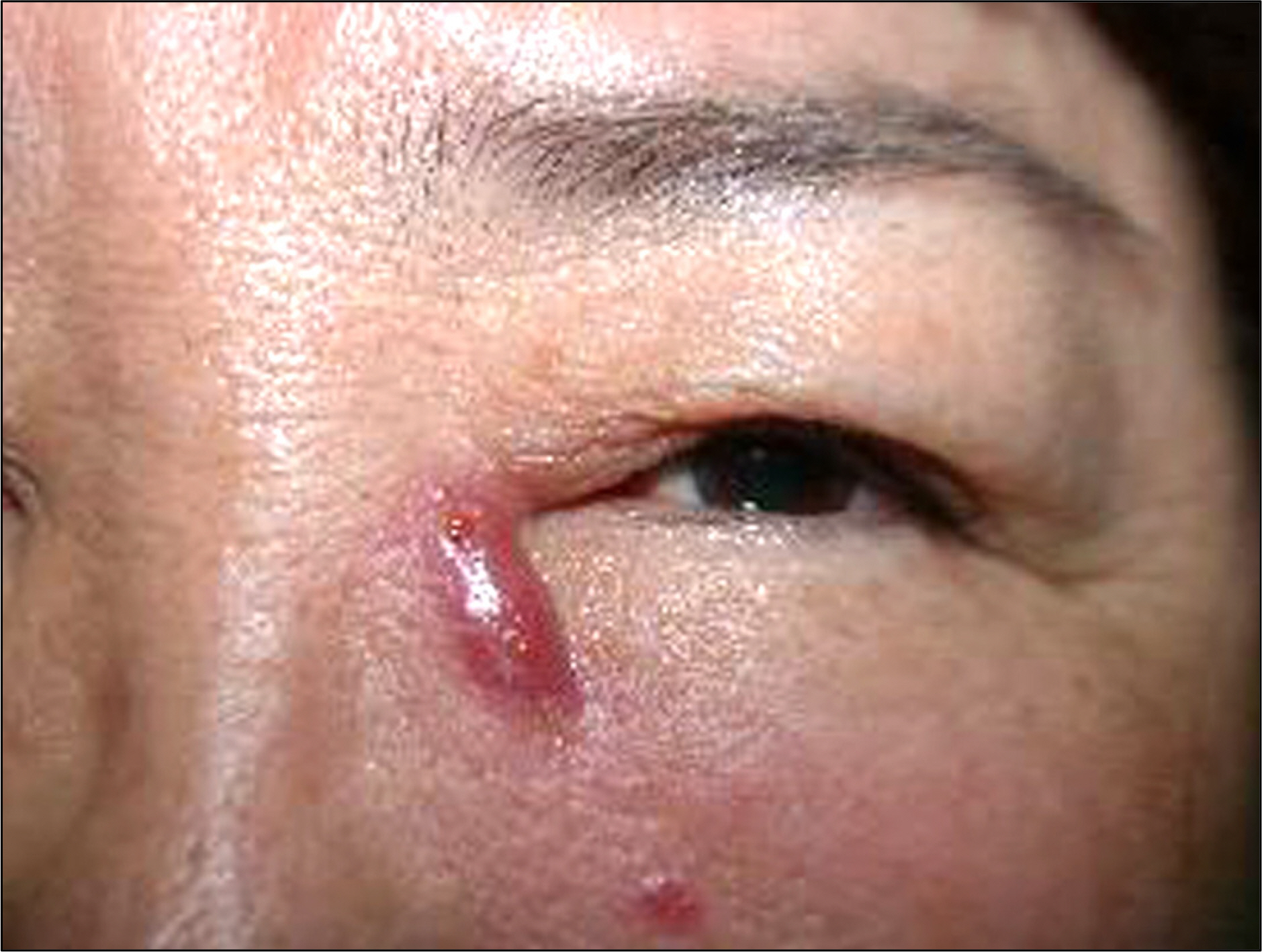
Figure 1. A 53-year-old woman with multiple elevated erythematous lesions 4 weeks after external dacryocystorhinostomy.
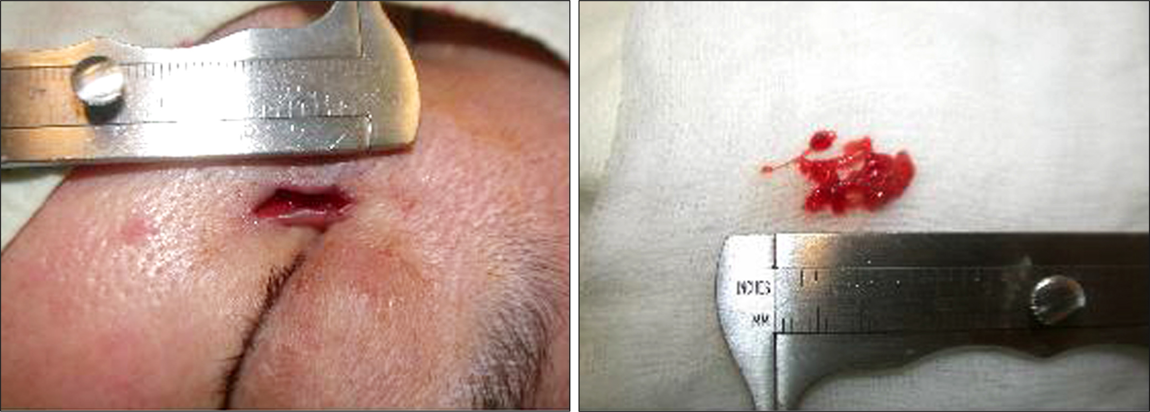
Figure 2. Excisional biopsy.
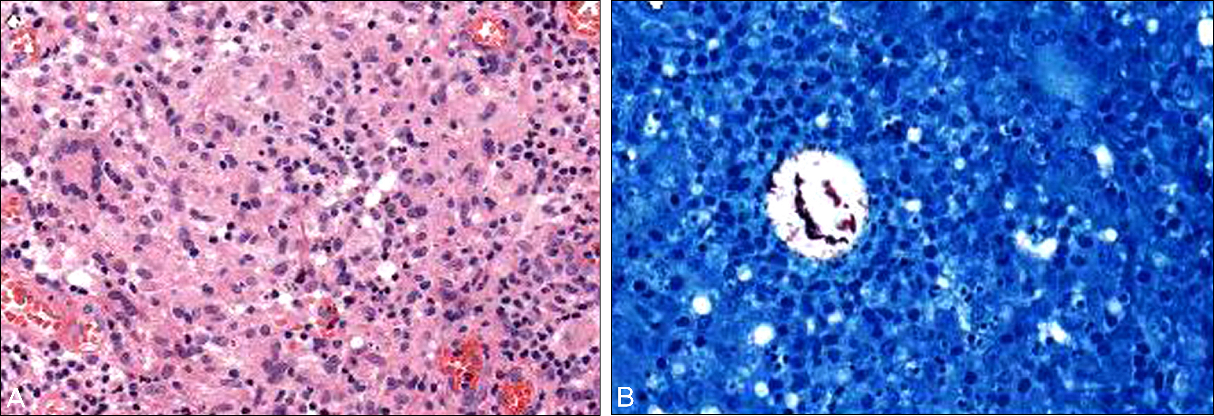
Figure 3. (A) Histopathologic examination showing granulomatous inflammation with areas of necrosis and a microabscess (stain, hematoxylin and eosin; magnification, ×400). (B) Acid-fast bacilli (stain, Ziehl Neelsen; magnification, ×400).
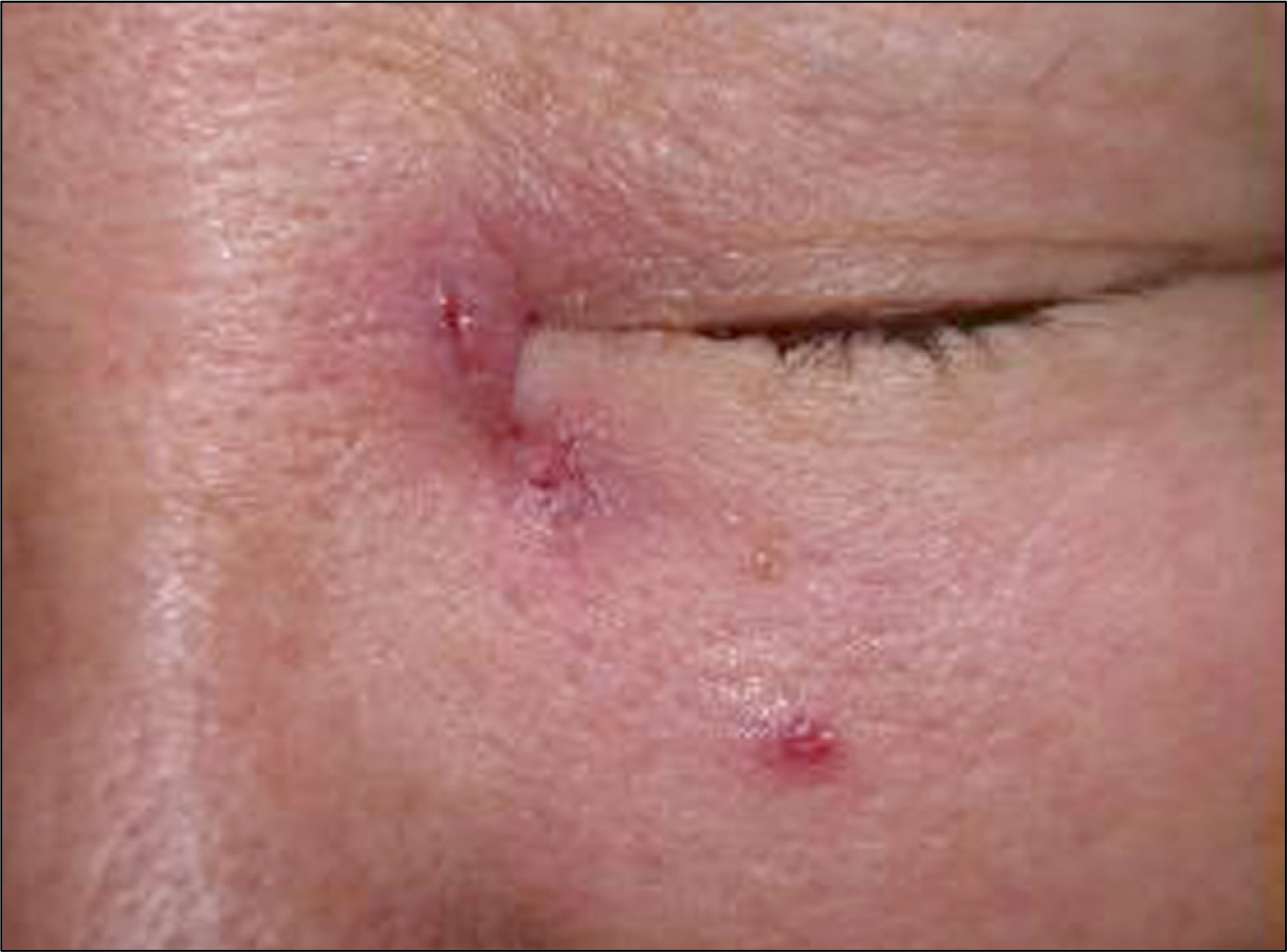
Figure 4. A decreased erythematous granulomatous lesions 2 months after excisional biopsy and anti-tuberculosis treatment.
Cited by 1 articles
-
Two Cases of Skin Necrosis after Canaliculodacryocystorhinostomy in Young Women
Eun Young Cho, Jun Hyuk Son
J Korean Ophthalmol Soc. 2015;56(11):1798-1803. doi: 10.3341/jkos.2015.56.11.1798.
Reference
-
References
1. Rosen N, Sharir M, Moverman DC, Rosner M. Dacryocystorhinostomy with silicone tubes: evaluation of 253 cases. Ophthalmic Surg. 1989; 20:115–9.
Article2. Safranek TJ, Jarvis WR, Carson LA, et al. Mycobacterium chelonae wound infections after plastic surgery employing contaminated gentian violet skin-marking solution. N Engl J Med. 1987; 317:197–201.3. Meisler DM, Friedlaender MH, Okumoto M. Mycobacterium chelonei keratitis. Am J Ophthalmol. 1982; 94:398–401.
Article4. Dugel PU, Holland GN, Brown HH, et al. Mycobacterium fortu-itum keratitis. Am J Ophthalmol. 1988; 105:661–9.
Article5. Smiddy WE, Miller D, Flynn HW Jr. Scleral buckle infections due to atypical mycobacteria. Retina. 1991; 11:394–8.
Article6. Smith RE, Salz JJ, Moors R, et al. Mycobacterium chelonei and orbital granuloma after tear duct probing. Am J Ophthalmol. 1980; 89:139–41.
Article7. Chang WJ, Tse DT, Rosa RH Jr, Miller D. Periocular atypical mycobacterial infections. Ophthalmology. 1999; 106:86–90.
Article8. Koh WJ, Kwon OJ, Lee KS. Diagnosis and treatment of nontuberculous mycobacterial pulmonary diseases: a Korean perspective. J Korean Med Sci. 2005; 20:913–25.
Article9. American Thoracic Society. Diagnosis and treatment of disease caused by nontuberculous mycobacteria. Am J Respir Crit Care Med. 1997; 156:S1–25.10. British Thoracic Society. Management of opportunistic mycobacterial infections: Joint Tuberculosis Committee Guidelines 1999. Thorax. 2000; 55:210–8.11. O'Brien RJ, Geiter LJ, Snider DE Jr. The epidemiology of nontuberculous mycobacterial diseases in the United States. Results from a national survey. Am Rev Respir Dis. 1987; 135:1007–14.12. Kubica GP, Baess I, Gordon RE, et al. A co-operative numerical analysis of rapidly growing mycobacteria. J Gen Microbiol. 1972; 73:55–70.13. Kusunoki S, Ezaki T. Proposal of Mycobacterium peregrinum sp. nov., nom. rev., and elevation of Mycobacterium chelonae subsp. abscessus (Kubica et al.) to species status: Mycobacterium abscessus comb. nov. Int J Syst Bacteriol. 1992; 42:240–5.
Article14. Ashford DA, Kellerman S, Yakrus M, et al. Pseudo-outbreak of septicemia due to rapidly growing mycobacteria associated with extrinsic contamination of culture supplement. J Clin Microbiol. 1997; 35:2040–2.
Article15. Koh WJ, Kwon OJ. Treatment of nontuberculous mycobacterial pulmonary disease. Tuberc Respir Dis. 2004; 56:5–17.16. Wallace RJ Jr, Brown BA, Griffith DE. Nosocomial outbreaks/ pseudo-outbreaks caused by nontuberculous mycobacteria. Annu Rev Microbiol. 1998; 52:453–90.
- Full Text Links
-
- Actions
-
Cited
- CITED
-
- Close
- Share
-
- Similar articles
-
- Delayed Nontuberculous Mycobacterium Manifestation 1 Year after a Dog Bite on the Hand
- Chronic Granulomatous Infection of Soft Tissue Complicated by Trauma of a Lower Leg
- A Case of Skin and Soft Tissue Infection by Mycobacterium massiliense
- Cutaneous Lesion due to Mycobacterium Fortuitum
- A Case of Nontuberculous Mycobacterium Infection Complicated by an Esophagomediastinal Fistula in a Human Immunodeficiency Virus Patient
