J Korean Ophthalmol Soc.
2011 Jan;52(1):107-111. 10.3341/jkos.2011.52.1.107.
Atypical Ocular and Optical Coherence Tomographic Findings With Presumed Miliary Tuberculosis
- Affiliations
-
- 1Department of Ophthalmology, Eul-ji University Hospital, Daejeon, Korea. snlee@eulji.ac.kr
- KMID: 2214146
- DOI: http://doi.org/10.3341/jkos.2011.52.1.107
Abstract
- PURPOSE
To report clinical features and optical coherence tomographic findings of presumed atypical ocular tuberculosis associated with tuberculosis lymphadenitis and encephalomeningitis.
CASE SUMMARY
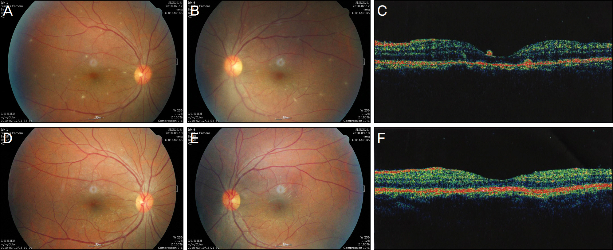
A 28-year-old female with lymphadenitis in the axillary area presented with a fever and headache of a one week duration. CSF study and MRI findings implied tuberculosis encephalomeningitis, and presumed tuberculosis uveitis manifested with visual disturbance after five days. Ocular symptoms were aggravated and showed anterior iridocyclitis, vitritis, macular edema, and multifocal retinitis with miliary granuloma that was distinct from choroiditis or typical tuberculosis granuloma. After the patient received anti-tuberculosis medication and systemic corticosteroids, significant improvements in visual acuity, ocular findings and OCT results were observed.
CONCLUSIONS
Ocular tuberculosis can present with various clinical findings, and caution should be taken so as not to misdiagnose based on these characteristics. In the present case, anti-tuberculosis medication and systemic steroids resulted in the resolution of inflammation. In such cases, monitoring the posterior pole lesion via OCT may be helpful in determining improvement.
MeSH Terms
Figure
Cited by 1 articles
-
The Clinical Manifestations and Differential Diagnosis of Tuberculosis Serpiginous-like Choroiditis and Serpiginous Choroiditis
Sung Hyun Ahn, Nam Chun Cho, Min Ahn, In Cheon You, Jin Gu Jeong
J Korean Ophthalmol Soc. 2017;58(1):50-55. doi: 10.3341/jkos.2017.58.1.50.
Reference
-
References
1. Tabbara KF. Tuberculosis. Curr Opin Ophthalmol. 2007; 18:493–501.
Article2. Sheu SJ, Shyu JS, Chen LM, et al. Ocular manifestations of tuberculosis. Ophthalmology. 2001; 108:1580–5.
Article3. Moon S, Son J, Chang W. A case of oculomotor nerve palsy and choroidal tuberculous granuloma associated with tuberculous meningoencephalitis. Korean J Ophthalmol. 2008; 22:201–4.
Article4. Vasconcelos-Santos DV, Zierhut M, Rao NA. Strengths and weak-nesses of diagnostic tools for tuberculous uveitis. Ocul Immunol Inflamm. 2009; 17:351–5.
Article5. Gupta A, Bansal R, Gupta V, et al. Ocular signs predictive of tubercular uveitis. Am J Ophthalmol. 2010; 149:562–70.
Article6. Suzuki J, Oh-I K, Kezuka T, et al. Comparison of patients with ocular tuberculosis in the 1990s and the 2000s. Jpn J Ophthalmol. 2010; 54:19–23.
Article7. Sinha MK, Garg RK, Anuradha HK, et al. Vision impairment in tuberculous meningitis: predictors and prognosis. J Neurol Sci. 2010; 15:27–32.
Article8. Mehta S. Fundus fluorescein angiography of choroidal tubercles: case reports and review of literature. Indian J Ophthalmol. 2006; 54:273–5.
Article9. Salman A, Parmar P, Rajamohan M, et al. Optical coherence tomography in choroidal tuberculosis. Am J Ophthalmol. 2006; 142:170–2.
Article10. Gupta V, Gupta A, Sachdeva N, et al. Successful management of tubercular subretinal granulomas. Ocul Immunol Inflamm. 2006; 14:35–40.
Article
- Full Text Links
- Actions
-
Cited
- CITED
-
- Close
- Share
- Similar articles
-
- Ocular manifestations of systemic tuberculosis: Report of 3 cases
- The Fundus in Tuberculosis in Children
- Optical Coherence Tomography Findings in Three Cases of Albinism
- A Case of Metastatic Tuberculosis Abscess Associated with Miliary Tuberculosis
- Can A Sudden Sensorineural Hearing Loss Occur Due to Miliary Tuberculosis?


