J Korean Surg Soc.
2011 May;80(5):348-354. 10.4174/jkss.2011.80.5.348.
Measurement of carotid artery stenosis: correlation analysis between B-mode ultrasonography and contrast arteriography
- Affiliations
-
- 1Division of Vascular Surgery, Department of Surgery, Samsung Medical Center, Sungkyunkwan University School of Medicine, Seoul, Korea. ywkim@skku.edu
- KMID: 2212160
- DOI: http://doi.org/10.4174/jkss.2011.80.5.348
Abstract
- PURPOSE
To evaluate the efficacy of B-mode ultrasonography (US) in measurement of carotid stenosis% (CS%).
METHODS
One hundred and thirth-three carotid arteries in 96 patients who underwent both carotid US and carotid arteriography (CA) were included in this retrospective study. To measure CS% on US, a cross sectional view of the most stenotic segment of the internal carotid artery was captured and residual diameter and original diameter of that segment were measured with electronic caliper on the same plane and in the same direction. To measure CS% on an angiogram, we used European Carotid Surgery Trial (ECST) and the North American Symptomatic Carotid Endarterectomy Trial (NASCET) methods. Pearson's correlation analysis and linear regression analysis were used to determine the correlation between CS% on an US and angiogram.
RESULTS
Pearson's correlation coefficient (R) between CS% measured in US and CA were 0.853 (ECST method, P < 0.001) and 0.828 (NASCET method, P < 0.001). Accuracies of B-mode US were 93.2%, 88.0%, and 81.2% for estimating CS% by ECST method and 86.5%, 82.7%, and 82% for estimating CS% by NASCET method.
CONCLUSION
CS% measured in B-mode US was simpler and showed a strong positive correlation with that measured on an arteriogram either ECST or NASCET method.
Keyword
MeSH Terms
Figure
-
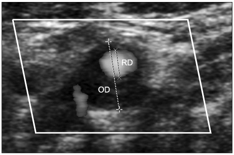
Fig. 1 Calculation of carotid stenosis% on a transverse scan color flow ultrasonography image of the internal carotid artery: residual diameter (RD) indicates the shortest diameter of residual lumen of the most stenotic segment of the internal carotid artery and original diameter (OD) indicates a diameter from the outer media-to-outer media in a same plane and a same direction with RD.
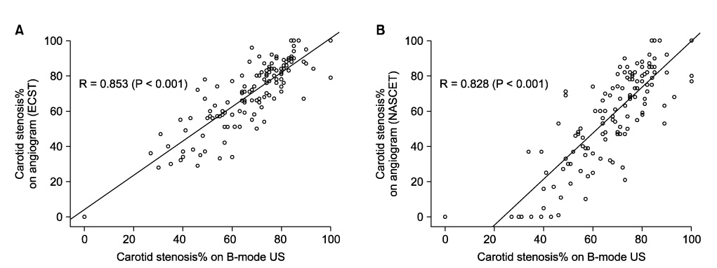
Fig. 2 Scatter diagrams of the carotid stenosis% (CS%) measured ultrasonography (US) in reference to CS% measured on carotid angiogram. (A) Angiographic CS% measured by European Carotid Surgery Trial method. (B) Angiographic CS% measured by North American Symptomatic Carotid Endarterectomy Trial method.
Reference
-
1. Hobson RW 2nd, Mackey WC, Ascher E, Murad MH, Calligaro KD, Comerota AJ, et al. Management of atherosclerotic carotid artery disease: clinical practice guidelines of the Society for Vascular Surgery. J Vasc Surg. 2008. 48:480–486.2. Grant EG, Benson CB, Moneta GL, Alexandrov AV, Baker JD, Bluth EI, et al. Carotid artery stenosis: gray-scale and Doppler US diagnosis--Society of Radiologists in Ultrasound Consensus Conference. Radiology. 2003. 229:340–346.3. Lemne C, Jogestrand T, de Faire U. Carotid intima-media thickness and plaque in borderline hypertension. Stroke. 1995. 26:34–39.4. Wendelhag I, Gustavsson T, Suurküla M, Berglund G, Wikstrand J. Ultrasound measurement of wall thickness in the carotid artery: fundamental principles and description of a computerized analysing system. Clin Physiol. 1991. 11:565–577.5. Executive Committee for the Asymptomatic Carotid Atherosclerosis Study. Endarterectomy for asymptomatic carotid artery stenosis. JAMA. 1995. 273:1421–1428.6. North American Symptomatic Carotid Endarterectomy Trial Collaborators. Beneficial effect of carotid endarterectomy in symptomatic patients with high-grade carotid stenosis. N Engl J Med. 1991. 325:445–453.7. Higashida RT, Meyers PM, Phatouros CC, Connors JJ 3rd, Barr JD, Sacks D, et al. Reporting standards for carotid artery angioplasty and stent placement. J Vasc Interv Radiol. 2004. 15:421–422.8. Corriveau MM, Johnston KW. Interobserver variability of carotid Doppler peak velocity measurements among technologists in an ICAVL-accredited vascular laboratory. J Vasc Surg. 2004. 39:735–741.9. Kuntz KM, Polak JF, Whittemore AD, Skillman JJ, Kent KC. Duplex ultrasound criteria for the identification of carotid stenosis should be laboratory specific. Stroke. 1997. 28:597–602.10. Mead GE, Lewis SC, Wardlaw JM. Variability in Doppler ultrasound influences referral of patients for carotid surgery. Eur J Ultrasound. 2000. 12:137–143.11. AbuRahma AF, Robinson PA, Strickler DL, Alberts S, Young L. Proposed new duplex classification for threshold stenoses used in various symptomatic and asymptomatic carotid endarterectomy trials. Ann Vasc Surg. 1998. 12:349–358.12. Hood DB, Mattos MA, Mansour A, Ramsey DE, Hodgson KJ, Barkmeier LD, et al. Prospective evaluation of new duplex criteria to identify 70% internal carotid artery stenosis. J Vasc Surg. 1996. 23:254–261.13. Moneta GL, Edwards JM, Papanicolaou G, Hatsukami T, Taylor LM Jr, Strandness DE Jr, et al. Screening for asymptomatic internal carotid artery stenosis: duplex criteria for discriminating 60% to 99% stenosis. J Vasc Surg. 1995. 21:989–994.14. Moneta GL, Edwards JM, Chitwood RW, Taylor LM Jr, Lee RW, Cummings CA, et al. Correlation of North American Symptomatic Carotid Endarterectomy Trial (NASCET) angiographic definition of 70% to 99% internal carotid artery stenosis with duplex scanning. J Vasc Surg. 1993. 17:152–157.15. Sprouse LR 2nd, Meier GH, Parent FN, Demasi RJ, Lesar CJ, Nelms C, et al. Are we undertreating carotid stenoses diagnosed by ultrasound alone? Vasc Endovascular Surg. 2005. 39:143–151.16. MacKenzie KS, French-Sherry E, Burns K, Pooley T, Bassiouny HS. B-mode ultrasound measurement of carotid bifurcation stenoses: is it reliable? Vasc Endovascular Surg. 2002. 36:123–135.17. Handa N, Matsumoto M, Maeda H, Hougaku H, Ogawa S, Fukunaga R, et al. Ultrasonic evaluation of early carotid atherosclerosis. Stroke. 1990. 21:1567–1572.18. O'Donnell TF Jr, Erdoes L, Mackey WC, McCullough J, Shepard A, Heggerick P, et al. Correlation of B-mode ultrasound imaging and arteriography with pathologic findings at carotid endarterectomy. Arch Surg. 1985. 120:443–449.19. Wolverson MK, Bashiti HM, Peterson GJ. Ultrasonic tissue characterization of atheromatous plaques using a high resolution real time scanner. Ultrasound Med Biol. 1983. 9:599–609.20. Rotstein AH, Gibson RN, King PM. Direct B-mode NASCET-style stenosis measurement and Doppler ultrasound as parameters for assessment of internal carotid artery stenosis. Australas Radiol. 2002. 46:52–56.21. Jmor S, El-Atrozy T, Griffin M, Tegos T, Dhanjil S, Nicolaides A. Grading internal carotid artery stenosis using B-mode ultrasound (in vivo study). Eur J Vasc Endovasc Surg. 1999. 18:315–322.22. Golledge J, Ellis M, Sabharwal T, Sikdar T, Davies AH, Greenhalgh RM. Selection of patients for carotid endarterectomy. J Vasc Surg. 1999. 30:122–130.23. Beebe HG, Salles-Cunha SX, Scissons RP, Dosick SM, Whalen RC, Gale SS, et al. Carotid arterial ultrasound scan imaging: a direct approach to stenosis measurement. J Vasc Surg. 1999. 29:838–844.
- Full Text Links
-
- Actions
-
Cited
- CITED
-
- Close
- Share
-
- Similar articles
-
- Relationship Between Carotid and Coronary Atherosclerosis in the Elderly
- Diagnostic vascular ultrasonography with the help of color Doppler and contrast-enhanced ultrasonography
- Comparison between conventional duplex ultrasonography and the dual-gate Doppler mode for hemodynamic measurements of the carotid arteries
- General principles of carotid Doppler ultrasonography
- Overestimation of Carotid Artery Stenosis by Carotid Duplex Ultrasonography Due to Contralateral Occlusion
