Artifacts in Retinal Nerve Fiber Layer Analysis Using Optical Coherence Tomography
- Affiliations
-
- 1Department of Ophthalmology, Korea University College of Medicine, Seoul, Korea.
- 2Department of Ophthalmology, Kyung Hee University College of Medicine, East-West Neo Medical Center, Seoul, Korea. hukang@dreamwiz.com
- KMID: 2211708
- DOI: http://doi.org/10.3341/jkos.2008.49.5.778
Abstract
-
PURPOSE: To investigate the frequency and characteristics of artifacts which can cause errors in retinal nerve fiber layer nalysis using optical coherence tomography (OCT).
METHODS
The frequency, characteristics and retinal lesions responsible for the artifact were analyzed for 179 patients (338 eyes) by OCT. All images were categorized into two groups according to the presence of artifacts and then, the differences between the two groups were analyzed by t-test and cross-tabulation analysis in terms of age, refractive error, peripapillary atrophic areas, and type of glaucoma.
RESULTS
The male to female ratio was 1.37:1, average age was 47.6+/-15.7 years and average degree of refraction was -1.78+/-0.23 diopter. Artifacts were noted in 64 eyes (18.9%), and were present in the temporal quadrant in 12 eyes (18.8%), superior quadrant in 51 eyes (79.7%), nasal quadrant in 19 eyes (87.5%), and inferior quadrant in 8 eyes (48.4%). The average angle of the artifact was 138 degrees. Although retinal lesion, age, and glaucoma type were not significantly different between the two groups, peripapillary atrophy and myopia were significantly more common in the group with the artifact.
CONCLUSIONS
When analyzing retinal nerve fiber layer by OCT, artifacts should be considered, especially in cases of peripapillary atrophy and myopia.
MeSH Terms
Figure
-
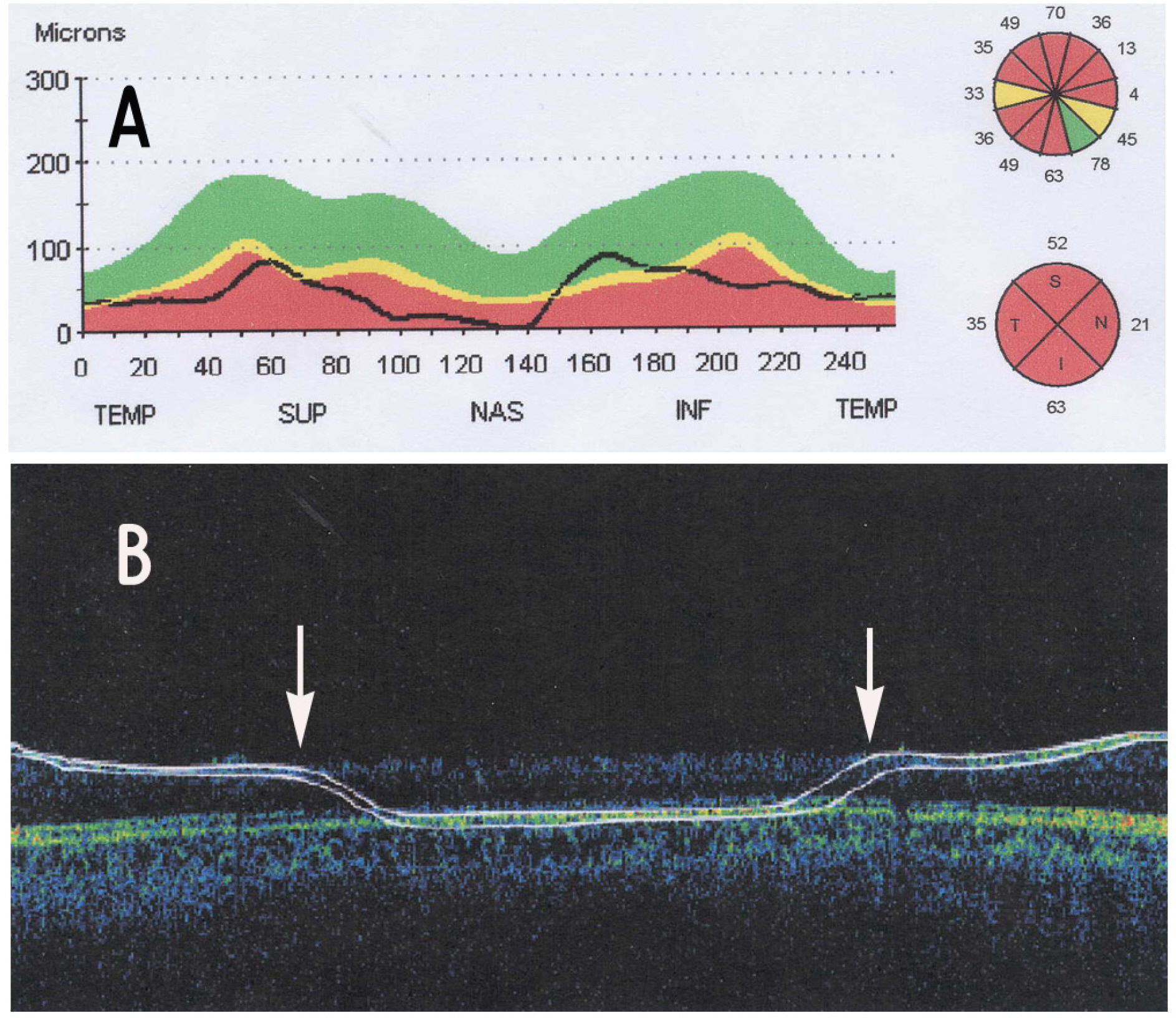
Figure 1. An example of artifact (area between two arrows) in retinal nerve fiber layer (RNFL) thickness analysis by optical coherence tomography. Fast RNFL thickness (3.4) scan mode (A) reveals generalized reduction of RNFL thickness for nearly 360 degree. However, RNFL thickness (2.27Xdisc) scan mode (B) shows incorrect nerve fiber layer detection in nasal quadrant expecially..

Figure 2. Visual field (A) and red-free retinal nerve fiber layer (RNFL) photogarphy (B) of the same patient in Figure 1. These results do not correlate with the RNFL defect area in artifact obtained by optical coherence tomography.
Cited by 1 articles
-
Retinal Nerve Fiber Layer Thickness in Children With Glaucoma
Kui Dong Kang, Aman Shah B. Abdul Majid, Yeon Deok Kim, Jee hyun Kwag, Hye Bin Yim
J Korean Ophthalmol Soc. 2009;50(6):887-892. doi: 10.3341/jkos.2009.50.6.887.
Reference
-
References
1. Sommer A, Pollak I, Naumence AE. Optic disc parameters and onset of glaucomatous field loss. I. Methods and progressive changes in disc morphology. Arch Ophthalmol. 1979; 97:1444–8.2. Quigley HA, Addicks EM. Green WR. Optic nerve damage in human glaucoma. III. Quantitative correlation of nerve fiber loss and visual field defects in glaucoma, ischemic neuropathy, papilledema and toxic neuropathy. Arch Ophthalmol. 1982; 100:135–46.3. Caprioli J, Miller JM, Sears M. Quantitative evaluation of the optic nerve head in patients with unilateral visual field loss from primary open angle glaucoma. Ophthalmology. 1987; 94:1484–7.4. Pederson JE, Anderson DR. The mode of progressive disc cupping in ocular hypertension and glaucoma. Arch Ophthalmol. 1998; 180:490–5.
Article5. Varma R, Steinmann WC, Scott IU. Expert agreement in evaluating the optic disc for glaucoma. Ophthalmology. 1992; 99:215–21.
Article6. Quigley HA, Kats J, Derick RJ, et al. An evaluation of optic disc and nerve fiber layer examinations in monitoring progression of early glaucoma damage. Ophthalmology. 1992; 99:19–28.
Article7. Caprioli J, Miller JM. Measurement of relative nerve fiber layer surface height in glaucoma. Ophthalmology. 1989; 96:633–41.8. Quigley HA, Addicks EM, Green WR. Optic nerve damage in human glaucoma. Arch Ophthalmol. 1982; 100:135–46.
Article9. Sommer A, Miller NR, Pollack I, et al. The nerve fiber layer in the diagnosis of glaucoma. Arch Ophthalmol. 1977; 95:2149–56.
Article10. Sommer A, Kats J, Quigley HA, et al. Clinically detectable nerve fiber atrophy precedes the onset of glaucomatous field loss. Arch Ophthalmol. 1991; 109:77–83.
Article11. Cho CH, Kee CW. Association of retinal nerve fiber layer thickness measured by optical coherence tomography and automatic perimetry. J Korean Ophthalmol Soc. 2002; 43:1032–9.12. Colen TP, Lemij HG. Motion artifacts in scanning polarimetry. Ophthalmology. 2002; 31:342–5.13. Pons ME, Rothman RF, Ozden RG, et al. Vitreous opacities affect scanning laser polarimetry measurement. Am J Ophthalmol. 2001; 131:511–3.14. Schuman JS, Pedut-Kloizman T, Hertzmark E, et al. Reproducibility of nerve fiber layer thickness measurements using optical coherence tomography. Opthalmology. 1996; 103:1889–98.
Article15. Sadda SR, Wu Z, Walsh AC, et al. Errors in retinal thickness measurements obtained by optical coherence tomography. Ophthalmology. 2006; 113:285–93.
Article16. Ray R, Stinnet SS, Jaffe GJ. Evaluation of image artifact produced by optical coherence tomography of retinal pathology. Am J Ophthalmol. 2005; 139:18–29.
Article17. Foos RY. Vitreoretinal juncture: topographical variation. Invest Ophthalmol. 1972; 11:801–8.18. Roth AM, Foos RY. Surface structure of the optic nerve head. Am J Ophthalmol. 1972; 74:977–85.
Article19. Stein DM, Wollstein G, Ishikawa H, et al. Effect of corneal drying on optical coherence tomography. Ophthalmology. 2006; 113:985–91.
Article20. Hee MR. Artifacts in optical coherence tomography topographic maps. Am J Ophthalmol. 2005; 139:154–5.
Article
- Full Text Links
-
- Actions
-
Cited
- CITED
-
- Close
- Share
-
- Similar articles
-
- Asymmetry Analysis of the Retinal Nerve Fiber Layer Thickness in Normal Eyes using Optical Coherence Tomography
- Thicknesses of the Fovea and Retinal Nerve Fiber Layer in Amblyopic and Normal Eyes in Children
- Thicknesses of Macular Retinal Layer and Peripapillary Retinal Nerve Fiber Layer in Patients with Hyperopic Anisometropic Amblyopia
- A Case of Retinal Herniation through Peripapillary Pit Resulting in Retinal Nerve Fiber Layer Defect
- Retinal Nerve Fiber Layer Thickness Measured by Spectral Domain Optical Coherence Tomography in Healthy Koreans
