Asymmetry Analysis of the Retinal Nerve Fiber Layer Thickness in Normal Eyes using Optical Coherence Tomography
- Affiliations
-
- 1Cheil Eye Hospital, Daegu, Korea. ophth@korea.com
- KMID: 754440
- DOI: http://doi.org/10.3341/kjo.2005.19.4.281
Abstract
- PURPOSE
To investigate the asymmetry of the retinal nerve fiber layer thickness (RNFLT) with respect to the horizontal and vertical meridian and between the right and left eye in normal subjects. METHODS: The RNFLT was measured in 121 normal volunteers by optical coherence tomography (OCT). The RNFLT was analyzed by dividing the circle scanning area (diameter 3.4 mm) around the optic disc into 4 quadrants and 12 sectors. RESULTS: There was a significant difference between the RNFLT of the nasal and temporal quadrant in individual eyes. There was a significant difference between the RNFLT of corresponding sectors with respect to the vertical or horizontal meridian in individual eyes. The nasal and temporal RNFLTs were asymmetrical between the right and left eye in the quadrant and sector analysis. The RNFLT of the nasal and temporal quadrant was thicker in the right eye. The nasal and inferior RNFLT measured by OCT had a significant correlation with degree of refractive error. CONCLUSIONS: In normal subjects without significant anisometropia, there was significant asymmetry of the RNFLT for each eye as well as between the right and left eye.
Keyword
MeSH Terms
Figure
-

Fig. 1 (A) Four 90-degree quadrants of retinal nerve fiber layer thickness measured by a circle scanning diameter of 3.4 mm around the optic disc by OCT in the right and left eyes. S: superior quadrant, I: inferior quadrant, N: nasal quadrant, T: temporal quadrant. (B) Twelve equal 30-degree sectors of retinal nerve fiber layer thickness measured by a circle scanning diameter of 3.4 mm around the optic disc by OCT in the right and left eye.
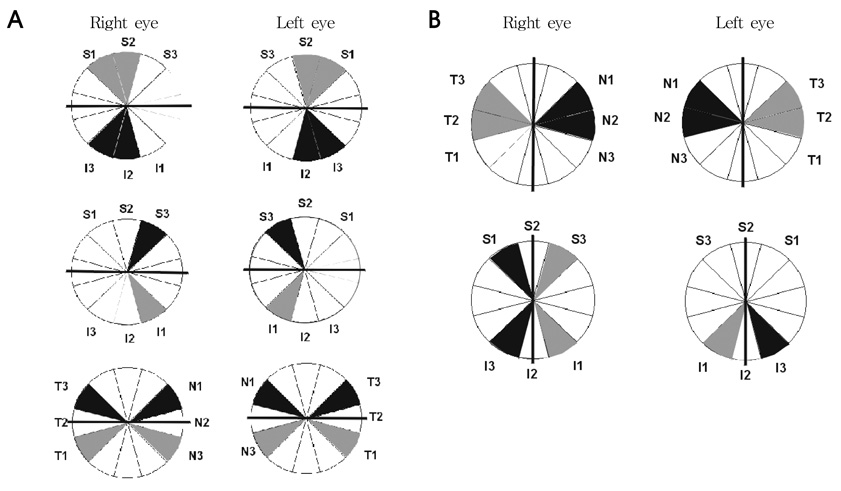
Fig. 2 (A) The difference of RNFLT between superior sectors and inferior sectors, symbolized by black and gray shading, respectively, was accepted as statistically significant (p<0.05). RNFLT in the black-colored sector was thicker than RNFLT in the gray-colored sector. Inferior sectors were thicker in those sectors corresponding to the arcuate area, but superior sectors were thicker in the non-arcuate area. (B) The difference of RNFLT between nasal sectors and temporal sectors, symbolized by the black and gray shading, respectively, was accepted as statistically significant (p<0.05). The RNFLT in the black colored sector was thicker than the RNFLT in gray colored sector. The RNFLT of nasal sectors close to the horizontal poles was thicker than that of temporal sectors. The RNFLT of temporal sectors close to the vertical meridian was thicker than that of nasal sectors.

Fig. 3 The difference in RNFLT between the right and left eye, symbolized by black and gray shading, respectively, was accepted as statistically significant (p<0.05). RNFLT in the black-colored sector was thicker than RNFLT in the gray-colored sector. RNFLT was thicker in the right than the left eye at the nasal and temporal sectors
Reference
-
1. Quigley HA, Dunkelberger GR, Green WR. Retinal ganglion cell atrophy correlated with automated pyrometry in human eyes with glaucoma. Am J Ophthalmol. 1989. 107:453–464.2. Quigley HA, Dunkelberger GR, Green WR. Chronic human glaucoma causing selectively greater loss of large optic nerve fibers. Ophthalmology. 1988. 95:357–363.3. Sommer A, Katz J, Quigley HA, et al. Clinically detectable nerve fiber atrophy precedes the onset of glaucomatous field loss. Arch Ophthalmol. 1991. 109:77–83.4. Iwata K. Ophthalmoscopy in the detection of optic disc and retinal nerve fiber layer changes in early glaucoma. Surv Ophthalmol. 1989. 33:447–448.5. Quigley HA, Miller NR, George T. Clinical evaluation of nerve fiber layer atrophy as an indicator of glaucomatous optic nerve damage. Arch Ophthalmol. 1980. 98:1564–1571.6. Sommer A, Miller NR, Pollack I, et al. The nerve fiber layer in the diagnosis of glaucoma. Arch Ophthalmol. 1977. 95:2149–2156.7. Yasuo K, Kaori M, Yumi K, et al. Asymmetries of the retinal nerve fibre layer thickness in normal eyes. Br J Ophthalmol. 2000. 84:469–472.8. Essock EA, Sinai MJ, Fechtner RD. Interocular symmetry in nerve fiber layer thickness of normal eyes as determined by polarimetry. J Glaucoma. 1999. 8:90–98.9. Schuman JS, Hee MR, Arya AV, et al. Optical coherence tomography: a new tool for glaucoma diagnosis. Curr Opin Ophthalmol. 1995. 6:89–95.10. Klemm M, Rumberger E, Walter A, Richard G. Reproducibility of measuring retinal nerve fiber density. Comparison of optical coherence tomography with the nerve fiber analyzer and the Heidelberg retinal tomography device. Ophthalmologe. 2002. 99:345–351.11. Armaly MF. Genetic determination of cup/disc ratio of the optic nerve. Arch ophthalmol. 1967. 78:5.12. Ong LS, Mitchell P, Healey PR, et al. Asymmetry in optic disc parameters: the Blue Mountains Eye Study. Invest Ophthalmol Vis Sci. 1999. 40:849.13. Weinreb RN, Shakiba S, Zangwill L. Scanning laser polarimetry to measure the nerve fiber layer of normal and glaucomatous eyes. Am J Ophthalmol. 1995. 119:627–636.14. Anton A, Zangwill L, Emdadi A, et al. Nerve fiber layer measurement with scanning laser polarimetry in ocular hypertension. Arch Ophthalmol. 1997. 115:331–334.15. Dichtl A, Jonas JB, Naumann GO. Retinal nerve fiber layer thickness in human eyes. Graefe's Arch Clin Exp Ophthalmol. 1999. 237:474–479.16. Balazsi AG, Rootman J, Drance SM, et al. The effect of age on the nerve fiber population of the human optic nerve. Am J Ophthalmol. 1984. 97:760–766.17. Mikelberg FS, Drance SM, Schulzer M, et al. The normal human optic nerve. Axon count and axon diameter distribution. Ophthalmology. 1989. 96:1325–1328.18. Repka MX, Quigley HA. The effect of age on normal human optic nerve fiber number and diameter. Ophthalmology. 1989. 96:26–32.
- Full Text Links
-
- Actions
-
Cited
- CITED
-
- Close
- Share
-
- Similar articles
-
- Thicknesses of the Fovea and Retinal Nerve Fiber Layer in Amblyopic and Normal Eyes in Children
- Correlation Between Central Corneal Thickness and Retinal Nerve Fiber Layer Thickness in Normal Tension Glaucoma
- Comparison of Time Domain OCT and Spectrum Domain OCT for Retinal Nerve Fiber Layer Assessment
- Retinal Nerve Fiber Layer Thickness Measurement Using Swept Source Optical Coherence Tomography in Healthy Korean
- Reproducibility of Retinal Nerve Fiber Layer Thickness Evaluation by Nerve Fiber Analyzer
