Second Reactivation of Neurocysticercosis: A Case Report
- Affiliations
-
- 1Department of Radiology, Gachon University, Gil Hospital, Incheon, Korea. h2y@gilhospital.com
- KMID: 2208880
- DOI: http://doi.org/10.3348/jksr.2010.63.1.15
Abstract
- This report describes the first case involving a second reactivation of neurocysticercosis. There was peripheral enhancement and surrounding edema at multiple calcified lesions in both cerebral hemispheres on the brain MRI. One must be aware of the possibility of reactivation of neurocysticercosis to make the correct diagnosis.
Figure
-
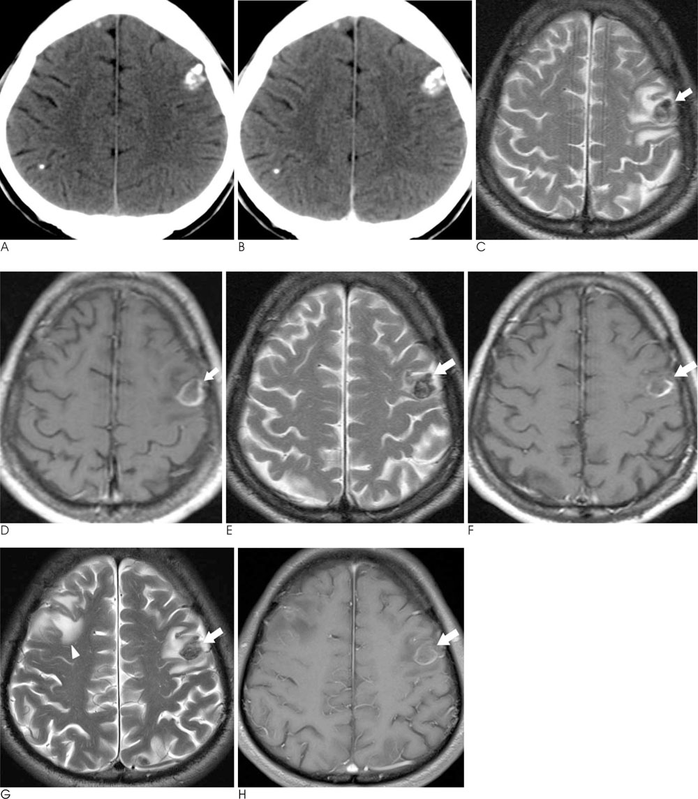
Fig. 1 A 59-year-old man with known neurocysticercosis. A, B. The pre- & post-contrast brain CT that was performed for routine follow-up before the first reactivation of neurocysticercosis. Multifocal dense nodular calcifications are detected in both cerebral hemispheres, and especially in the left frontal lobe (A). There are no abnormal enhancing lesions around the nodular calcifications on post-contrast brain CT (B). C, D. The brain MRI with gadolinium enhancement that was performed in 2002 at the first reactivation of neurocysticercosis. A dark signal intensity lesion is shown in the left frontal lobe on the T2-weighted magnetic resonance image (arrow on C). This lesion is compatible with the dense nodular calcifications seen on the previous brain CT (A & B). There is edema around the lesion. A diffuse thick band of enhancement is noted in the peripheral portion of the lesion on the contrast-enhanced T1-weighted magnetic resonance image (arrow on D). E, F. The brain MRI with gadolinium enhancement that was performed in 2004 during routine follow-up and the patient had neurologic symptoms. An unchanged dark signal intensity lesion is shown in the left frontal lobe on the T2-weighted magnetic resonance image (E). Edema around the lesion, which was detected on the previous brain MRI, is not shown. A diffuse thin band of enhancement is noted at the peripheral portion of the lesion on the contrast-enhanced T1-weighted magnetic resonance image (F). G, H. The brain MRI with gadolinium enhancement that was performed at presentation at the time of the last admission. Edema is noted again around the unchanged dark signal intensity lesion in the left frontal lobe on the T2-weighted magnetic resonance image (G). Additional edema is visible around the nodular calcification (not shown) in the right frontal lobe (arrowhead). A diffuse thin band of enhancement is not changed at the peripheral portion of the lesion on the contrast-enhanced T1-weighted magnetic resonance image since the previous study in 2004 (H).
Reference
-
1. White AC Jr. Neurocysticercosis: Updates on epidemiology, pathogenesis, diagnosis, and management. Annu Rev Med. 2000; 51:187–206.2. Kong Y, Cho SY, Cho MS, Kwon OS, Kang WS. Seroepidemiological observation of taenia solium cysticercosis in epileptic patients in Korea. J Korean Med Sci. 1993; 8:145–152.3. Escobar A. The pathology of neurocysticercosis. In : Palacios E, Rodriguez-Carbajal J, Taveras JM, editors. Cysticercosis of the central nervous system. Springfield, III: Charles C. Thomas;1983. p. 27–54.4. Hawk MW, Shahlaie K, Kim KD, Theis JH. Neurocysticercosis: a review. Surg Neurol. 2005; 63:123–132.5. Garcia HH, Del Brutto OH. Imaging findings in neurocysticercosis. Acta Trop. 2003; 87:71–78.6. Sheth TN, Pillon L, Keystone J, Kucharczyk W. Persistent MR contrast enhancement of calcified neurocysticercosis lesions. AJNR Am J Neuroradiol. 1998; 19:79–82.7. Sheth TN, Lee C, Kucharczyk W, Keystone J. Reactivation of neurocysticercosis: case report. Am J Trop Med Hyg. 1999; 60:664–667.
- Full Text Links
-
- Actions
-
Cited
- CITED
-
- Close
- Share
-
- Similar articles
-
- Calcified Neurocysticercosis that Invaded the Subarachnoid Space Presenting as Focal Status Epilepticus
- Extraparenchymal (Racemose) Neurocysticercosis and Its Multitude Manifestations: A Comprehensive Review
- Neurocysticercosis Presenting as Homonymous Hemianopia
- A Case of Neurocysticercosis in Entire Spinal Level
- Stereotactic Surgery of Neurocysticercosis
