Prevalence of Disc Degeneration in Asymptomatic Korean Subjects. Part 2 : Cervical Spine
- Affiliations
-
- 1Department of Neurosurgery, 21st Century Hospital, Seoul, Korea.
- 2Department of Neurosurgery, Ewha Womans University School of Medicine, Mokdong Hospital, Seoul, Korea. sjkimmd@unitel.co.kr
- 3Department of Radiology, Ewha Womans University School of Medicine, Mokdong Hospital, Seoul, Korea.
- KMID: 2190684
- DOI: http://doi.org/10.3340/jkns.2013.53.2.89
Abstract
OBJECTIVE
Similar to back pain, neck pain has recently shown to have increasing prevalence. Magnetic resonance imaging (MRI) is useful in identifying the causes of neck pain. However, MRI shows not only pathological lesions but also physiological changes at the same time, and there are few Korean data. The authors have attempted to analyze the prevalence of disc degeneration in highly selective asymptomatic Korean subjects using MRI.
METHODS
We performed 3 T MRI sagittal scans from C2 to T1 on 102 asymptomatic subjects (50 men and 52 women) who visited our hospital between the ages of 14 and 82 years (mean age 46.3 years). All images were read independently by three observers (two neurosurgeons and one neuroradiologist) who were not given any information about the subjects. We classified grading for cervical disc herniation (HN), annular fissure (AF), and nucleus degeneration (ND), using disc degeneration classification.
RESULTS
The prevalence of HN, AF, and ND were 81.0%, 85.9%, and 95.4%, respectively. High prevalence of HN, AF, and ND was shown compared to previous literature.
CONCLUSION
In asymptomatic Korean subjects, the abnormal findings of 3 T MRI showed a high prevalence in HN, AF, and ND. Several factors might play important roles in these results, such as population-specific characters, MRI field strength, and disc degeneration grading system.
MeSH Terms
Figure
-

Fig. 1 References of image for disc degeneration are shown. The criteria of classification are described in Table 2 and 3. A : Herniation. G0, normal. G1, diffuse bulging. G2, protrusion. G3, extrusion. B : Annular fissure. G0, normal. G1, Annular fissure at posterior or posterolateral site. G2, annular fissure at anterior site. G3, annular fissure at anterior and posterior site. C : Nucleus degeneration. G0, normal. G1, normal disc height and hyperintense nucleus signal with horizontal dark band. G2, decreased disc height and hyperintense nucleus signal with or without horizontal dark band. G3, normal disc height and decreased nucleus signal with slightly or heterogeneous irregularity. G4, slightly decreased disc height and decreased nucleus signal with slightly or heterogeneous irregularity. G5, moderately decreased disc height and decreased nucleus signal with moderately. G6, collapsed disc height and hypointense nucleus signal.
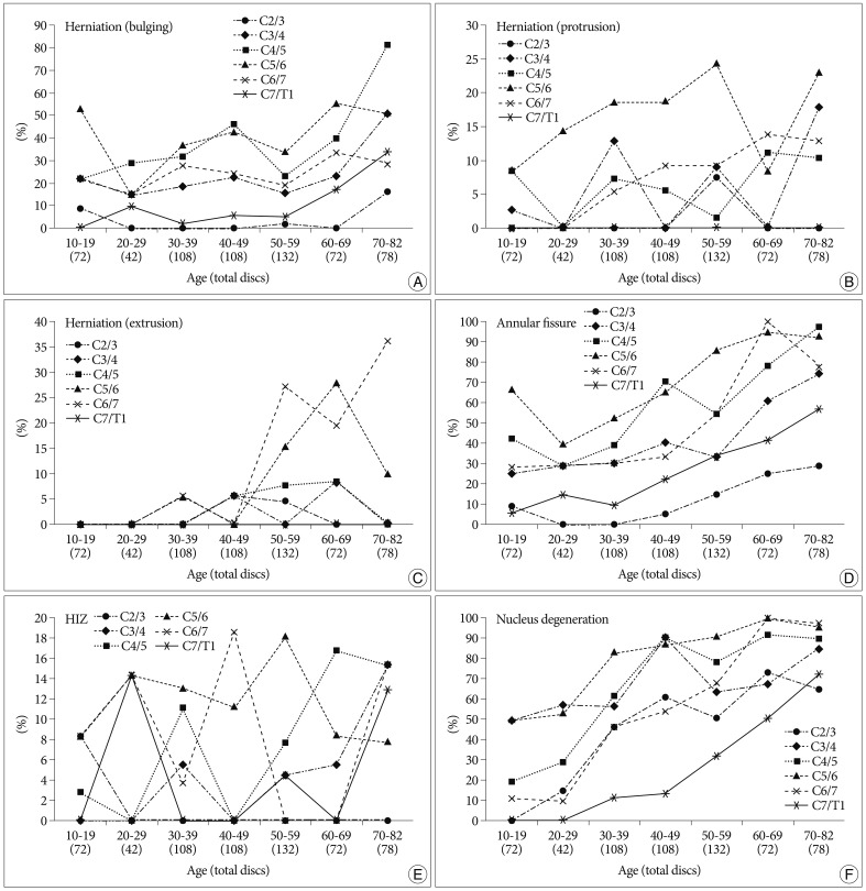
Fig. 2 Graph of disc degeneration prevalence according to age and level, calculated by disc count. A, B and C : Herniation. D : Annular fissure. E : High-signal intensity zone (HIZ). F : Nucleus degeneration.
Cited by 2 articles
-
Nonoperative interventions for spinal pain
Dong Ah Shin, Hyoung Ihl Kim
J Korean Med Assoc. 2014;57(4):308-317. doi: 10.5124/jkma.2014.57.4.308.Prevalence of Disc Degeneration in Asymptomatic Korean Subjects. Part 3 : Cervical and Lumbar Relationship
Sang Jin Kim, Tae Hoon Lee, Seong Yi
J Korean Neurosurg Soc. 2013;53(3):167-173. doi: 10.3340/jkns.2013.53.3.167.
Reference
-
1. Aprill C, Bogduk N. High-intensity zone : a diagnostic sign of painful lumbar disc on magnetic resonance imaging. Br J Radiol. 1992; 65:361–369. PMID: 1535257.
Article2. Boden SD, McCowin PR, Davis DO, Dina TS, Mark AS, Wiesel S. Abnormal magnetic-resonance scans of the cervical spine in asymptomatic subjects. A prospective investigation. J Bone Joint Surg Am. 1990; 72:1178–1184. PMID: 2398088.
Article3. Boos N, Rieder R, Schade V, Spratt KF, Semmer N, Aebi M. 1995 Volvo Award in clinical sciences. The diagnostic accuracy of magnetic resonance imaging, work perception, and psychosocial factors in identifying symptomatic disc herniations. Spine (Phila Pa 1976). 1995; 20:2613–2625. PMID: 8747239.
Article4. Brant-Zawadzki MN, Jensen MC, Obuchowski N, Ross JS, Modic MT. Interobserver and intraobserver variability in interpretation of lumbar disc abnormalities. A comparison of two nomenclatures. Spine (Phila Pa 1976). 1995; 20:1257–1263. discussion 1264. PMID: 7660234.
Article5. Christe A, Läubli R, Guzman R, Berlemann U, Moore RJ, Schroth G, et al. Degeneration of the cervical disc : histology compared with radiography and magnetic resonance imaging. Neuroradiology. 2005; 47:721–729. PMID: 16136264.
Article6. Fejer R, Kyvik KO, Hartvigsen J. The prevalence of neck pain in the world population : a systematic critical review of the literature. Eur Spine J. 2006; 15:834–848. PMID: 15999284.
Article7. Fries P, Runge VM, Kirchin MA, Watkins DM, Buecker A, Schneider G. Magnetic resonance imaging of the spine at 3 Tesla. Semin Musculoskelet Radiol. 2008; 12:238–252. PMID: 18850504.
Article8. Griffith JF, Wang YX, Antonio GE, Choi KC, Yu A, Ahuja AT, et al. Modified Pfirrmann grading system for lumbar intervertebral disc degeneration. Spine (Phila Pa 1976). 2007; 32:E708–E712. PMID: 18007231.
Article9. Harris RI, Macnab I. Structural changes in the lumbar intervertebral discs; their relationship to low back pain and sciatica. J Bone Joint Surg Br. 1954; 36-B:304–322. PMID: 13163117.10. Masaryk TJ, Ross JS, Modic MT, Boumphrey F, Bohlman H, Wilber G. High-resolution MR imaging of sequestered lumbar intervertebral disks. AJR Am J Roentgenol. 1988; 150:1155–1162. PMID: 3258720.
Article11. Matsumoto M, Fujimura Y, Suzuki N, Nishi Y, Nakamura M, Yabe Y, et al. MRI of cervical intervertebral discs in asymptomatic subjects. J Bone Joint Surg Br. 1998; 80:19–24. PMID: 9460946.
Article
- Full Text Links
-
- Actions
-
Cited
- CITED
-
- Close
- Share
-
- Similar articles
-
- Prevalence of Disc Degeneration in Asymptomatic Korean Subjects. Part 1 : Lumbar Spine
- Prevalence of Disc Degeneration in Asymptomatic Korean Subjects. Part 3 : Cervical and Lumbar Relationship
- Correlation of Cervical Disc Degeneration with Sagittal Alignments of Cervical Spine
- Clinical Relationship of Degenerative Changes between the Cervical and Lumbar Spine
- Comparison of Disc Degeneration between the Cervical and Lumbar Spine
