Comparison of Ultrasonography and Magnetic Resonance Imaging in Measurement of Lumbar Spine Anatomic Structures
- Affiliations
-
- 1Department of Orthopedic Surgery, School of Medicine, Wonkwang University, Iksan, Korea. osktg@wonkwang.ac.kr
- KMID: 2185390
- DOI: http://doi.org/10.4055/jkoa.2012.47.2.140
Abstract
- PURPOSE
The aim of this study was to determine the usefulness of ultrasonography for lumbar anatomical structure measurement.
MATERIALS AND METHODS
From January 2011 to April 2011, 41 patients (22 males, 19 females) with back pain who visited the outpatient department and underwent lumbar magnetic resonance imaging (MRI) were selected. In each level of L4 and L5, we measured the longest distance and horizontal distance between each inferior articular process based off a spinous process. We also measured the distance between the spinous process tip and the vertebral body posterior surface and the thickness and width of the multifidus muscle. All distances were measured with ultrasonography and MRI and the two measurement results were compared.
RESULTS
Using ultrasonography and MRI, we measured the distance between the spinous process tip and the posterior surface of the body. The distances were 39.16+/-8.71 mm/39.53+/-6.01 mm at L4 and 38.32+/-9.66 mm/37.74+/-10.54 mm at L5. The right multifidus muscle thickness measurements were 32.13+/-10.79 mm/33.84+/-9 mm at L4 and 31.32+/-10.04 mm/32.84+/-12.28 mm at L5. The measuring distance between the spinous process center to the posterior vertebral body surface and thickness of multifidus muscles by ultrasonography and MRI had significant correlations (p<0.05).
CONCLUSION
Limitations still exist in measuring the structure of lumbar anatomy with ultrasonography. However, measuring the distance between the spinous process center to the vertebral body posterior surface and multifidus muscle thickness was effective.
Keyword
MeSH Terms
Figure
-
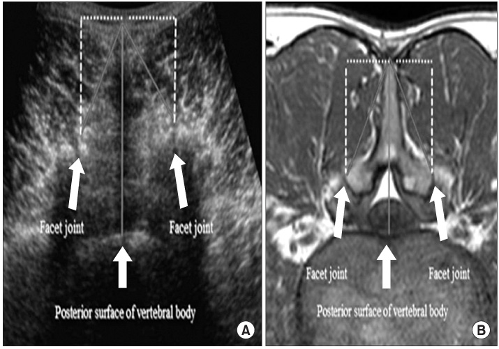
Figure 1 (A) Longest distance of inferior articular surface (oblique line) and horizontal distance of inferior articular surface (...) at axial transverse ultrasound image of L4-5 level. (B) Longest distance of inferior articular surface (oblique line) and horizontal distance of inferior articular surface (...) at axial transverse magnetic resonance imaging image of L4-5 level.
Cited by 2 articles
-
Comparison of Ultrasonography and Magnetic Resonance Imaging in Measurement of Lumbar Muscles
Chang Su Kim, Young Ha Woo, Dae Moo Shim, Tae Kyun Kim, Jeong Mi Lee, Bong Jun Jang, Byung Min Yoo
J Korean Orthop Assoc. 2016;51(5):371-377. doi: 10.4055/jkoa.2016.51.5.371.Usefulness of Ultrasound-guided Facet Joint Block in the Out Patient Clinics
Suk Joong Lee, Dae Moo Shim, Chang Su Kim, Sung Kyun Oh, Jae Seon Hwang
J Korean Soc Spine Surg. 2012;19(4):164-170. doi: 10.1874/JKSS.2021.19.4.164.
Reference
-
1. Kirchmair L, Entner T, Wissel J, Moriggl B, Kapral S, Mitterschiffthaler G. A study of the paravertebral anatomy for ultrasound-guided posterior lumbar plexus block. Anesth Analg. 2001. 93:477–481.
Article2. Stiffler KA, Jwayyed S, Wilber ST, Robinson A. The use of ultrasound to identify pertinent landmarks for lumbar puncture. Am J Emerg Med. 2007. 25:331–334.
Article3. Galiano K, Obwegeser AA, Bodner G, et al. Ultrasound guidance for facet joint injections in the lumbar spine: a computed tomography-controlled feasibility study. Anesth Analg. 2005. 101:579–583.
Article4. Shim DM, Kim TK, Lee SJ, Song SY. Comparison of ultrasonography and MRI in measuring of cervical soft tissue structure. J Korean Orthop Assoc. 2011. 46:282–287.
Article5. Stokes M, Rankin G, Newham DJ. Ultrasound imaging of lumbar multifidus muscle: normal reference ranges for measurements and practical guidance on the technique. Man Ther. 2005. 10:116–126.
Article6. Macintosh JE, Valencia F, Bogduk N, Munro RR. The morphology of the lumbar multifidus muscles. Clin Biomech. 1986. 1:196–204.7. Hashimoto BE, Kramer DJ, Wiitala L. Applications of musculoskeletal sonography. J Clin Ultrasound. 1999. 27:293–318.
Article8. Stoked M, Hides J, Nassiri D. Musculoskeletal ultrasound imaging: diagnostic and treatment aid in rehabilitation. Physical Theraphy reviews. 1997. 2:73–92.9. Park SK, Shim JI, Chung IW. Measurement of the oblique diameter of the lumbar spinal canal in Korean army-aged group by echographic method. J Korean Orthop Assoc. 1982. 17:763–771.10. Iannotti JP, Ciccone J, Buss DD, et al. Accuracy of office-based ultrasonography of the shoulder for the diagnosis of rotator cuff tears. J Bone Joint Surg Am. 2005. 87:1305–1311.
Article11. Bogduk N, Macintosh JE, Pearcy MJ. A universal model of the lumbar back muscles in the upright position. Spine (Phila Pa 1976). 1992. 17:897–913.
Article12. Hides JA, Cooper DH, Stokes MJ. Diagnostic ultrasound imaging for measurement of the lumbar multifidus muscle in normal young adults. Physiother Theory and Pract. 1992. 8:19–26.
Article13. Kader DF, Wardlaw D, Smith FW. Correlation between the MRI changes in the lumbar multifidus muscles and leg pain. Clin Radiol. 2000. 55:145–149.
Article14. Weber BR, Grob D, Dvorák J, Müntener M. Posterior surgical approach to the lumbar spine and its effect on the multifidus muscle. Spine (Phila Pa 1976). 1997. 22:1765–1772.
Article15. Zhao WP, Kawaguchi Y, Matsui H, Kanamori M, Kimura T. Histochemistry and morphology of the multifidus muscle in lumbar disc herniation: comparative study between diseased and normal sides. Spine (Phila Pa 1976). 2000. 25:2191–2199.16. Hides JA, Richardson CA, Jull GA. Multifidus muscle recovery is not automatic after resolution of acute, first-episode low back pain. Spine (Phila Pa 1976). 1996. 21:2763–2769.
Article17. Jesus FMR, Ferreira PH, Ferreira ML. Ultrasonographic measurement of neck muscle recruitment: a preliminary investigation. J Man Manip Ther. 2008. 16:89–92.
Article18. Comerford M, Mottram S. Stability dysfunction and low back pain. The Journal of Orthopaedic Medicine. 1998. 20:13–18.19. Lee JP, Tseng WY, Shau YW, Wang CL, Wang HK, Wang SF. Measurement of segmental cervical multifidus contraction by ultrasonography in asymptomatic adults. Man Ther. 2007. 12:286–294.
Article20. Hides JA, Richardson CA, Jull GA. Magnetic resonance imaging and ultrasonography of the lumbar multifidus muscle. Comparison of two different modalities. Spine (Phila Pa 1976). 1995. 20:54–58.21. Arzola C, Davies S, Rofaeel A, Carvalho JC. Ultrasound using the transverse approach to the lumbar spine provides reliable landmarks for labor epidurals. Anesth Analg. 2007. 104:1188–1192.
Article
- Full Text Links
-
- Actions
-
Cited
- CITED
-
- Close
- Share
-
- Similar articles
-
- Corrigendum: Comparison of Ultrasonography and Magnetic Resonance Imaging in Measurement of Lumbar Spine Anatomic Structures
- Utility of Limited Protocol Magnetic Resonance Imaging Lumbar Spine for Nerve Root Compression in a Developing Country, Is It Accurate and Cost Effective?
- Intradiscal Herniation of the Common Iliac Vessels: A Case Report
- MR Imaging and Ultrasonographic Findings of Tensor Fasciae Suralis Muscle: A Case Report
- Response to: “T1 Slope in the Cervical Spine Magnetic Resonance Imaging: A Novel Conceptâ€
