Intradiscal Herniation of the Common Iliac Vessels: A Case Report
- Affiliations
-
- 1Department of Radiology, Kyung Hee University Medical Center, Kyung Hee University College of Medicine, Seoul, Korea. t2star@khu.ac.kr
- 2Department of Radiology, Kyung Hee University Hospital at Gangdong, Kyung Hee University College of Medicine, Seoul, Korea.
- KMID: 2002944
- DOI: http://doi.org/10.3348/jksr.2011.65.4.411
Abstract
- In previously published spine related articles, common iliac vessel injuries have only been mentioned for complications resulting from a lumbar spine surgery. We present a case report of common iliac vessels herniating into a lumbar intervertebral disc incidentally found on magnetic resonance imaging and computed tomography angiography of the lumbar spine.
MeSH Terms
Figure
-
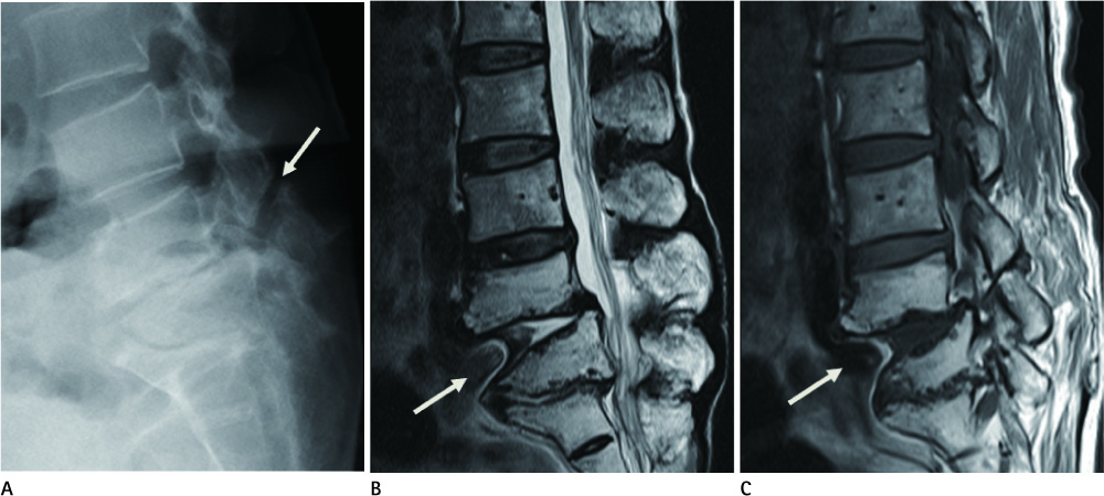
Fig. 1 Lateral plain radiograph (A) shows isthmic spondylolisthesis and severe disc degeneration of L4-L5 with bilateral pars interarticularis defects at L4 (arrow). Severe disc degeneration with retrolisthesis at L5-S1 and anterior wedged deformity of L5 body were seen. Midsagittal T2-weighted image (B) demonstrates herniation of the left common iliac vein and surrounding retroperitoneal fat into the L4-L5 disc (arrow). Central spinal canal narrowing at the level of L5 is seen with retrolisthesis of the L5-S1. Right parasagittal T1-weighted image (C) reveals herniation of the right common iliac artery and surrounding retroperitoneal fat into the L4-L5 disc.
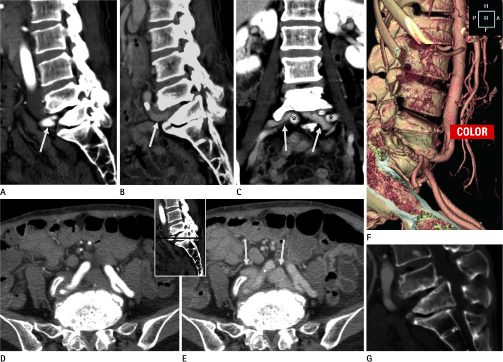
Fig. 2 Sagittal reformatted CT images show intradiscal herniation of the right common iliac artery bifurcation on a right parasagittal plane (A) and left common iliac vein on left parasagittal plane (B) at L4-L5. Coronal reformatted CT image (C), axial CT arterial phase image (D) and axial CT venous phase image (E) demonstrate the atypical location of both common iliac veins (arrows) and common iliac arteries (asterisks) just beneath the L4 body. Volume rendering CT angiography image of aorta and common iliac artery (F) shows intradiscal herniation of the right common iliac artery bifurcation. Sagittal reformatted CT image at the bone window setting (G) shows disc degeneration with vacuum disc phenomena, disc height loss and sclerosis of the bodies at L4-L5 with spondylolisthesis and L5-S1 with retrolisthesis, and the anterior wedged deformity of L5 body.
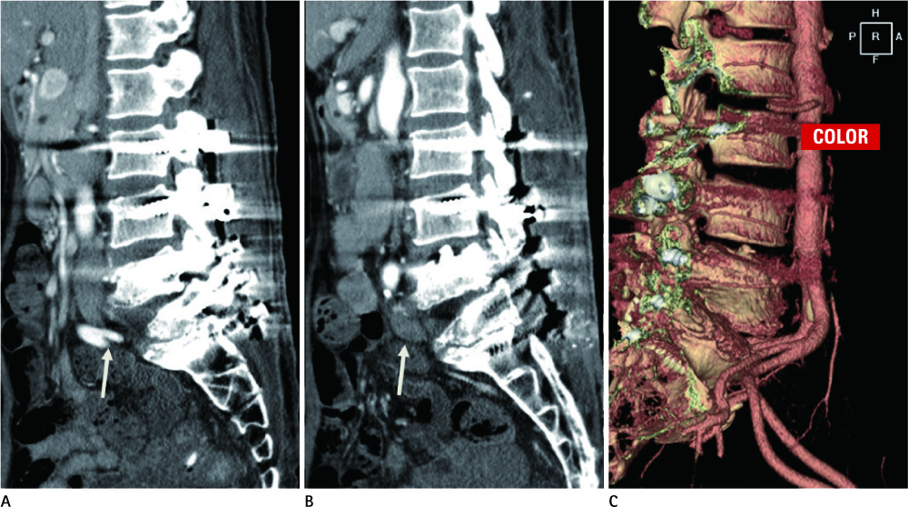
Fig. 3 Postoperative sagittal reformatted CT angiography arterial phase image (A) shows right common iliac artery bifurcation located at the retroperitoneum anterior to L4-S1. Postoperative sagittal reformatted CT angiography venous phase image (B) reveals the left common iliac vein located at the retroperitoneum anterior to discs L4-S1. There is no more intradiscal herniation of common iliac vessels. Postoperative volume rendering CT angiography image of aorta and common iliac artery (C) demonstrates right common iliac artery located anterior to L4-S1.
Reference
-
1. Samudrala S, Khoo LT, Rhim SC, Fessler RG. Complications during anterior surgery of the lumbar spine: an anatomically based study and review. Neurosurg Focus. 1999; 7:e9.2. Nam TK, Park SW, Shim HJ, Hwang SN. Endovascular treatment for common iliac artery injury complicating lumbar disc surgery: limited usefulness of temporary balloon occlusion. J Korean Neurosurg Soc. 2009; 46:261–264.3. Kang BU, Lee SH, Jeon SH, Park JD, Maeng DH, Choi YG, et al. An evaluation of vascular anatomy for minilaparotomic anterior L4-5 procedures. J Neurosurg Spine. 2006; 5:508–513.4. Devereaux MW. Anatomy and examination of the spine. Neurol Clin. 2007; 25:331–351.5. Hey HW, Hee HT. Lumbar degenerative spinal deformity: surgical options of PLIF, TLIF and MI-TLIF. Indian J Orthop. 2010; 44:159–162.6. Shih PY, Lau HP, Jeng CS, Hung MH, Chan KC, Cheng YJ. Iatrogenic left internal iliac artery perforation during lumbar discectomy. Acta Anaesthesiol Taiwan. 2009; 47:196–199.7. Goodkin R, Laska LL. Vascular and visceral injuries associated with lumbar disc surgery: medicolegal implications. Surg Neurol. 1998; 49:358–370. discussion 370-372.
- Full Text Links
-
- Actions
-
Cited
- CITED
-
- Close
- Share
-
- Similar articles
-
- Correlation Between Disk Morphology and Intradiscal Pressure in Lumbar Intervertebral Disk
- Lumbar Epidural Venography in the Diagnosis of Lumbar Disc Herniation
- Massive retroperitoneal hemorrhage due to a tearing of common iliac vein complicating lumbar diskectomy: a case report
- A Rare Case of Lumbar Traumatic Intradiscal Hematoma Followed by Repeatative Occupation Related Minor Trauma
- Multiple venous variations at the abdominopelvic region: a case report
