MR Imaging and Ultrasonographic Findings of Tensor Fasciae Suralis Muscle: A Case Report
- Affiliations
-
- 1Department of Radiology, Seoul Paik Hospital, Inje University College of Medicine, Seoul, Korea. jcshim96@unitel.co.kr
- KMID: 2068715
- DOI: http://doi.org/10.3348/jksr.2015.73.4.249
Abstract
- The tensor fasciae suralis muscle is a very rare anomalous muscle located in the popliteal region. This anatomic variation has been reported often through cadaver studies. However, there are only a few radiologic reports of this entity. We presented a case of tensor fasciae suralis muscle detected as an incidental finding in magnetic resonance imaging and ultrasound.
MeSH Terms
Figure
-
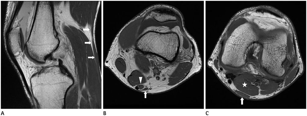
Fig. 1 MR images of the right knee. A. Sagittal proton density-weighted image shows an anomalous muscle (arrows) running superficially along the popliteal region. B, C. Axial T1-weighted images show an anomalous muscle (arrow) originating from the lateral aspect of semitendinosus muscle (arrowhead). Inferiorly, this muscle located posterior to the gastrocnemius medial head (asterisk). MR = magnetic resonance
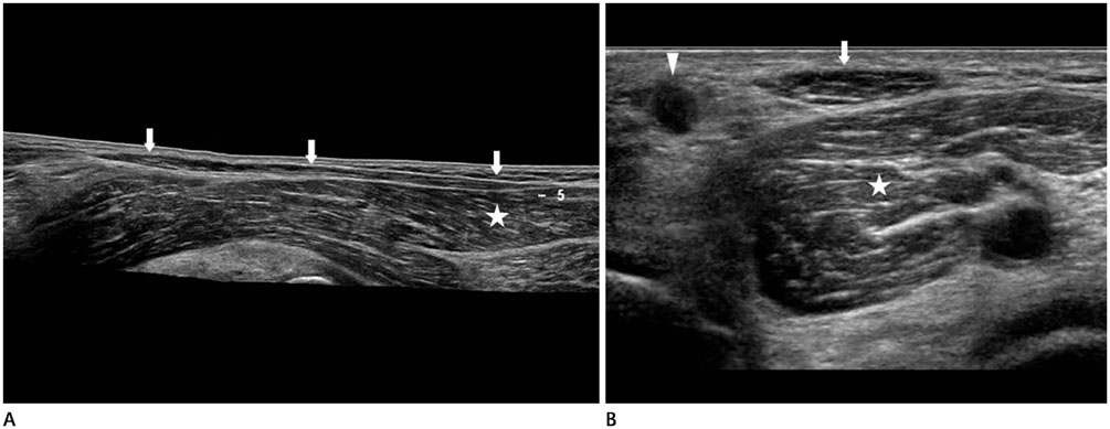
Fig. 2 US of the right popliteal region. A. Longitudinal panoramic US image shows the tensor fasciae suralis muscle (arrows) extending inferiorly and attaching to the gastocnemius medial head (asterisk). B. Transverse US image shows this muscle (arrow) located posterior to the gastrocnemius medial head (asterisk) and lateral to the semitendinosus tendon (arrowhead). It shows characteristic internal echotexture of the normal muscle. US = ultrasound
Cited by 1 articles
-
Clinical importance of tensor fasciae suralis arising from linea aspera along with short head of biceps femoris: a rare anomaly
Bincy M. George, Satheesha B. Nayak, Sapna Marpalli
Anat Cell Biol. 2019;52(1):90-92. doi: 10.5115/acb.2019.52.1.90.
Reference
-
1. Luca C, Stan C, Popescu D, Mota O, Popescu P, Davalciuc O. The tensor fasciae suralis muscle - case report. Revista Romana de Anatomie Functionala si Clinica, Macro- si Microscopica si de Antropologie. 2009; 8:501–503.2. Chason DP, Schultz SM, Fleckenstein JL. Tensor fasciae suralis: depiction on MR images. AJR Am J Roentgenol. 1995; 165:1220–1221.3. Montet X, Sandoz A, Mauget D, Martinoli C, Bianchi S. Sonographic and MRI appearance of tensor fasciae suralis muscle, an uncommon cause of popliteal swelling. Skeletal Radiol. 2002; 31:536–538.4. Sookur PA, Naraghi AM, Bleakney RR, Jalan R, Chan O, White LM. Accessory muscles: anatomy, symptoms, and radiologic evaluation. Radiographics. 2008; 28:481–499.5. Dunn AW. Anomalous muscles simulating soft-tissue tumors in the lower extremities. Report of three cases. J Bone Joint Surg Am. 1965; 47:1397–1400.6. Tubbs RS, Salter EG, Oakes WJ. Dissection of a rare accessory muscle of the leg: the tensor fasciae suralis muscle. Clin Anat. 2006; 19:571–572.7. Stoane JM, Gordon DH. MRI of an accessory semimembranosus muscle. J Comput Assist Tomogr. 1995; 19:161–162.
- Full Text Links
-
- Actions
-
Cited
- CITED
-
- Close
- Share
-
- Similar articles
-
- Clinical importance of tensor fasciae suralis arising from linea aspera along with short head of biceps femoris: a rare anomaly
- Anatomical and Radiological Study of the Vascular Distribution and Skin Territory for the Tensor Fasciae Latae Free Flap
- Morphometric Characteristics of Arterial Supply of Tensor Fasciae Latae Muscle in Korean
- Ultrasound and MR Imaging Findings of Vulvar Leiomyoma: Case Report
- Brain Diffusion Tensor MR Imaging
