Comparison of Closing-Wedge and Opening-Wedge High Tibial Osteotomies
- Affiliations
-
- 1Department of Orthopaedic Surgery, Gyeongsang National University School of Medicine, Jinju, Korea. shcho@gnu.kr
- KMID: 2185385
- DOI: http://doi.org/10.4055/jkoa.2012.47.2.104
Abstract
- PURPOSE
The aim of this study is to compare the clinical results and radiologic changes of closing-wedge high tibial osteotomy (CW HTO) and opening-wedge high tibial osteotomy (OW HTO).
MATERIALS AND METHODS
Seventy five knees were treated with a CW HTO and 41 with an OW HTO. For each patient the hip-knee-ankle (H-K-A) axis was evaluated and the degree of the medial compartmental arthrosis was measured by Ahlback-type radiological classification. The function of the knee was evaluated by the Tegner activity score, the Lysholm knee scoring scale and the Western Ontario and McMaster University index. Lateral radiographs were taken to assess the patellar height and the posterior tibial inclination.
RESULTS
In both groups significant improvement of the visual analogue scale and range of motion was achieved. The frontal plane H-K-A axis was corrected significantly from varus to the range of physiological valgus and the arthrosis of the medial compartment of the knee progressed gradually. The body mass index was significantly influential to the progression of arthrosis. The functions of the knee were improved significantly in all cases. In the closing-wedge group, the patella height was increased at the postoperative period, while it was decreased in the opening-wedge group. There was a tendency of a decrease of the tibial inclination in the CW HTO group and a statistically significant increase of the tibial inclination in the OW HTO group. Recurrence of varus occurred in sixteen cases.
CONCLUSION
In both groups, improvement of the function of the knee was achieved, but there was no statistical difference. However, the opposite result was found in the patella height and the tibial inclination.
Keyword
MeSH Terms
Figure
-

Figure 1 Visual analogue scale score. Preop, preoperative; Postop, postoperative; CWO, closing-wedge osteotomy; OWO, opening-wedge osteotomy.

Figure 2 Preoperative and postoperative standing radiographs showing satisfactory correction of the varus deformity and the fixation of the osteotomy with 90 degree angled blade plate designed by Se-Hyun Cho. The final standing x-ray shows maintenance of the correction after removal of the hardware.
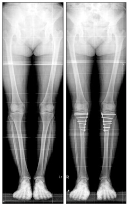
Figure 3 Preoperative varus gonarthrosis is well corrected by open wedge high tibial osteotomy fixed with anatomical plate.
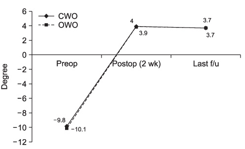
Figure 4 Correction in the frontal plane hip-knee-ankle axis. CWO, closing-wedge osteotomy; OWO, opening-wedge osteotomy; Preop, preoperative; Postop, postoperative; f/u, follow-up.
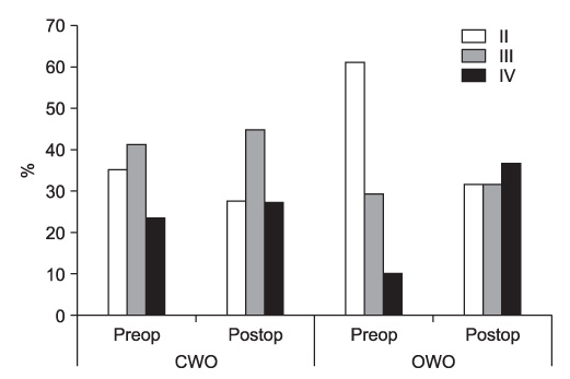
Figure 5 Radiologic changes evaluated by Ahlbäck classification. CWO, closing-wedge osteotomy; OWO, opening-wedge osteotomy; Preop, preoperative; Postop, postoperative.
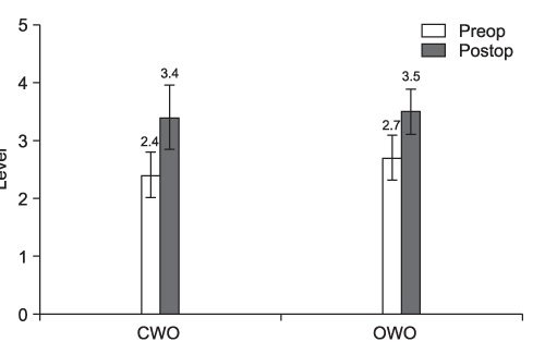
Figure 6 Tegner activity score. Preop, preoperative; Postop, postoperative; CWO, closing-wedge osteotomy; OWO, opening-wedge osteotomy.
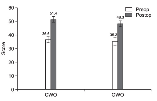
Figure 7 Lysholm knee scoring scale. Preop, preoperative; Postop, postoperative; CWO, closing-wedge osteotomy; OWO, opening-wedge osteotomy.
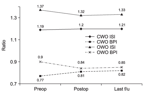
Figure 8 Patella height. CWO, closing-wedge osteotomy; ISI, Insall-Salvati Index; BPI, Blackburne Peel Index; OWO, opening-wedge osteotomy; Preop, preoperative; Postop, postoperative; f/u, follow-up.
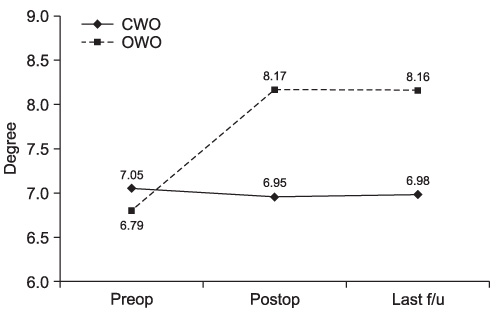
Figure 9 Posterior slope. CWO, closing-wedge osteotomy; OWO, opening-wedge osteotomy; Preop, preoperative; Postop, postoperative; f/u, follow-up.
Cited by 1 articles
-
Survival and Risk Factor Analysis of Open Wedge Tibial Osteotomy for Medial Unicompartmental Osteoarthritis
Gun Woo Kim, Eun Kyoo Song
J Korean Orthop Assoc. 2014;49(6):439-445. doi: 10.4055/jkoa.2014.49.6.439.
Reference
-
1. Aglietti P, Buzzi R, Vena LM, Baldini A, Mondaini A. High tibial valgus osteotomy for medial gonarthrosis: a 10- to 21-year study. J Knee Surg. 2003. 16:21–26.2. Cho SH, Park JT, Kim DH, Hwang SC. Conversion of total knee arthroplasty after high tibial osteotomy. J Korean Knee Soc. 2008. 20:38–43.3. Kesmezacar H, Erginer R, Ogut T, Seyahi A, Babacan M, Tenekecioglu Y. Evaluation of patellar height and measurement methods after valgus high tibial osteotomy. Knee Surg Sports Traumatol Arthrosc. 2005. 13:539–544.
Article4. Ozalay M, Ozkoc G, Circi E, et al. The correlation of correction magnitude and tibial slope changes following open wedge high tibial osteotomy. Knee Surg Sports Traumatol Arthrosc. 2008. 16:948–951.
Article5. Ward SR, Powers CM. The influence of patella alta on patellofemoral joint stress during normal and fast walking. Clin Biomech (Bristol, Avon). 2004. 19:1040–1047.
Article6. Ward SR, Terk MR, Powers CM. Patella alta: association with patellofemoral alignment and changes in contact area during weight-bearing. J Bone Joint Surg Am. 2007. 89:1749–1755.7. Fujisawa Y, Masuhara K, Shiomi S. The effect of high tibial osteotomy on osteoarthritis of the knee. An arthroscopic study of 54 knee joints. Orthop Clin North Am. 1979. 10:585–608.8. Weidow J, Cederlund CG, Ranstam J, Kärrholm J. Ahlbäck grading of osteoarthritis of the knee: poor reproducibility and validity based on visual inspection of the joint. Acta Orthop. 2006. 77:262–266.
Article9. Hoell S, Suttmoeller J, Stoll V, Fuchs S, Gosheger G. The high tibial osteotomy, open versus closed wedge, a comparison of methods in 108 patients. Arch Orthop Trauma Surg. 2005. 125:638–643.
Article10. Blackburne JS, Peel TE. A new method of measuring patellar height. J Bone Joint Surg Br. 1977. 59:241–242.
Article11. Insall J, Salvati E. Patella position in the normal knee joint. Radiology. 1971. 101:101–104.
Article12. Moore TM, Harvey JP Jr. Roentgenographic measurement of tibial-plateau depression due to fracture. J Bone Joint Surg Am. 1974. 56:155–160.
Article13. Haddad FS, Bentley G. Total knee arthroplasty after high tibial osteotomy: a medium-term review. J Arthroplasty. 2000. 15:597–603.14. Coventry MB, Ilstrup DM, Wallrichs SL. Proximal tibial osteotomy. A critical long-term study of eighty-seven cases. J Bone Joint Surg Am. 1993. 75:196–201.
Article15. Sprenger TR, Doerzbacher JF. Tibial osteotomy for the treatment of varus gonarthrosis. Survival and failure analysis to twenty-two years. J Bone Joint Surg Am. 2003. 85-A:469–474.16. Coventry MB. Osteotomy of the upper portion of the tibia for degenerative arthritis of the knee. A preliminary report. J Bone Joint Surg Am. 1965. 47:984–990.17. El-Azab H, Glabgly P, Paul J, Imhoff AB, Hinterwimmer S. Patellar height and posterior tibial slope after open- and closed-wedge high tibial osteotomy: a radiological study on 100 patients. Am J Sports Med. 2010. 38:323–329.18. Brouwer RW, Bierma-Zeinstra SM, van Koeveringe AJ, Verhaar JA. Patellar height and the inclination of the tibial plateau after high tibial osteotomy. The open versus the closed-wedge technique. J Bone Joint Surg Br. 2005. 87:1227–1232.19. Giagounidis EM, Sell S. High tibial osteotomy: factors influencing the duration of satisfactory function. Arch Orthop Trauma Surg. 1999. 119:445–449.
Article20. Cho SH, Hwang SC, Park JS, Lee SH. Change of the patellar height and tibial inclination after opening- and closing-wedge high tibial osteotomy. J Korean Knee Soc. 2010. 22:193–199.21. Scuderi GR, Windsor RE, Insall JN. Observations on patellar height after proximal tibial osteotomy. J Bone Joint Surg Am. 1989. 71:245–248.
Article22. Seil R, Müller B, Georg T, Kohn D, Rupp S. Reliability and interobserver variability in radiological patellar height ratios. Knee Surg Sports Traumatol Arthrosc. 2000. 8:231–236.
Article23. Tigani D, Ferrari D, Trentani P, Barbanti-Brodano G, Trentani F. Patellar height after high tibial osteotomy. Int Orthop. 2001. 24:331–334.
Article24. Noyes FR, Goebel SX, West J. Opening wedge tibial osteotomy: the 3-triangle method to correct axial alignment and tibial slope. Am J Sports Med. 2005. 33:378–387.
Article25. Wright JM, Heavrin B, Begg M, Sakyrd G, Sterett W. Observations on patellar height following opening wedge proximal tibial osteotomy. Am J Knee Surg. 2001. 14:163–173.26. Staubli AE, De Simoni C, Babst R, Lobenhoffer P. TomoFix: a new LCP-concept for open wedge osteotomy of the medial proximal tibia--early results in 92 cases. Injury. 2003. 34:Suppl 2. B55–B62.
- Full Text Links
-
- Actions
-
Cited
- CITED
-
- Close
- Share
-
- Similar articles
-
- Severe Genu Recurvatum after a Closing-wedge High Tibial Osteotomy: A Case Report
- High Tibial Osteotomy
- Comparison of High Tibial Osteotomy: Opening versus Closing Wedge Osteotomy
- Basic Principles and Current Trends of Medial Opening-Wedge High Tibial Osteotomy
- Delayed Onset of the Popliteal Artery Pseudoaneurysm Following Medial Opening Wedge High Tibial Osteotomy
