A Comparison of Different Techniques of Two-Dimensional Speckle-Tracking Strain Measurements of Right Ventricular Systolic Function in Patients with Acute Pulmonary Embolism
- Affiliations
-
- 1Department of Cardiology in Internal Medicine, School of Medicine, Chungnam National University, Chungnam National University Hospital, Daejeon, Korea. jaehpark@cnu.ac.kr
- 2Department of Vascular Surgery, School of Medicine, Chungnam National University, Chungnam National University Hospital, Daejeon, Korea.
- KMID: 2177461
- DOI: http://doi.org/10.4250/jcu.2014.22.2.65
Abstract
- BACKGROUND
Speckle-tracking echocardiography has been applied to measure right ventricular (RV) systolic function in various diseases. However, variations in strain measurement by different vendors have limited the application of these techniques for assessment of RV function. We sought to compare two methods for the assessment of RV systolic function in patients with acute pulmonary embolism (PE).
METHODS
From August 2007 to May 2011, all consecutive PE patients were prospectively included in this cohort study. Global longitudinal strains of RV measured with EchoPAC PC software (GLSRV-EchoPAC; GE Medical Systems) and velocity vector imaging (GLSRV-VVI; Siemens Medical Systems) were recorded on the same set of echocardiographic images.
RESULTS
We analyzed a total of 50 patients (12 males, 68 +/- 14 years) with acute PE in this study. GLSRV-EchoPAC and GLSRV-VVI were correlated (r = 0.793, p < 0.001) and they showed significant correlations with conventional echocardiographic parameters of RV systolic function and Log B-type natriuretic peptide (BNP) level. However, GLSRV-VVI only showed significant correlations with cardiac biomarkers as serum creatinine kinase-MB (r = 0.367, p = 0.010) and tropoinin-I concentrations (r = 0.294, p = 0.040).
CONCLUSION
GLSRV-VVI and GLSRV-EchoPAC showed significant correlations with conventional echocardiographic parameters of RV systolic function and LogBNP value in patients with PE.
MeSH Terms
Figure
-

Fig. 1 Correlation between the global longitudinal strain of the right ventricle (GLSRV) values by velocity vector imaging (GLSRV-VVI) and automated function imaging (GLSRV-EchoPAC). They show good correlation (A: scatter plot, B: Bland-Altman plot).
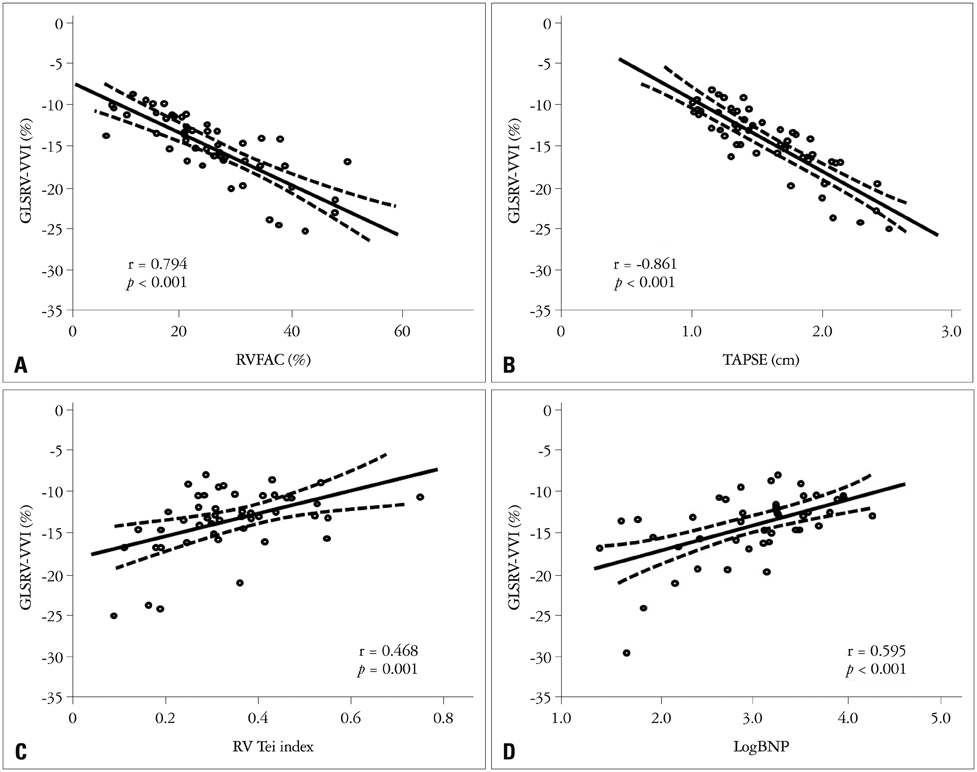
Fig. 2 Correlations between the global longitudinal strain of the right ventricle by velocity vector imaging (GLSRV-VVI) and echocardiographic parameters and serum B-natriuretic peptide (BNP) level. GLSRV-VVI shows good correlations with an RV fractional area change (RVFAC, A), tricuspid annular plane systolic excursion (TAPSE, B), RV Tei index (C), and LogBNP (D).

Fig. 3 Correlations between the global longitudinal strain of the right ventricle by automated function imaging (GLSRV-EchoPAC), echocardiographic parameters, and serum B-natriuretic peptide (BNP) level. GLSRV-VVI shows good correlations with an RV fractional area change (RVFAC, A), tricuspid annular plane systolic excursion (TAPSE, B), RV Tei index (C), and LogBNP (D).
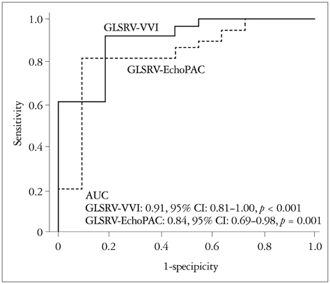
Fig. 4 Receiver operating curve analysis in the detection of right ventricular (RV) systolic dysfunction (determined by an RV fractional area change < 35%). The global longitudinal strain of the right ventricle by velocity vector imaging (GLSRV-VVI) shows a larger area under the curve than does the global longitudinal strain of the right ventricle by automatic function image (GLSRV-EchoPAC). However, there is no statistical significance [difference = 0.07, 95% confidence interval (CI): -0.03-0.17, p = 0.188] in the detection of RV systolic dysfunction.
Cited by 2 articles
-
Two-dimensional Echocardiographic Assessment of Myocardial Strain: Important Echocardiographic Parameter Readily Useful in Clinical Field
Jae-Hyeong Park
Korean Circ J. 2019;49(10):908-931. doi: 10.4070/kcj.2019.0200.Strain Analysis of the Right Ventricle Using Two-dimensional Echocardiography
Ju-Hee Lee, Jae-Hyeong Park
J Cardiovasc Imaging. 2018;26(3):111-124. doi: 10.4250/jcvi.2018.26.e11.
Reference
-
1. Torbicki A, Perrier A, Konstantinides S, Agnelli G, Galiè N, Pruszczyk P, Bengel F, Brady AJ, Ferreira D, Janssens U, Klepetko W, Mayer E, Remy-Jardin M, Bassand JP. ESC Committee for Practice Guidelines (CPG). Guidelines on the diagnosis and management of acute pulmonary embolism: the Task Force for the Diagnosis and Management of Acute Pulmonary Embolism of the European Society of Cardiology (ESC). Eur Heart J. 2008; 29:2276–2315.2. Kucher N, Rossi E, De Rosa M, Goldhaber SZ. Prognostic role of echocardiography among patients with acute pulmonary embolism and a systolic arterial pressure of 90 mm Hg or higher. Arch Intern Med. 2005; 165:1777–1781.
Article3. Grifoni S, Olivotto I, Cecchini P, Pieralli F, Camaiti A, Santoro G, Conti A, Agnelli G, Berni G. Short-term clinical outcome of patients with acute pulmonary embolism, normal blood pressure, and echocardiographic right ventricular dysfunction. Circulation. 2000; 101:2817–2822.
Article4. Grapsa J, Dawson D, Nihoyannopoulos P. Assessment of right ventricular structure and function in pulmonary hypertension. J Cardiovasc Ultrasound. 2011; 19:115–125.
Article5. Marwick TH. Measurement of strain and strain rate by echocardiography: ready for prime time? J Am Coll Cardiol. 2006; 47:1313–1327.6. Jamal F, Bergerot C, Argaud L, Loufouat J, Ovize M. Longitudinal strain quantitates regional right ventricular contractile function. Am J Physiol Heart Circ Physiol. 2003; 285:H2842–H2847.
Article7. Tandri H, Daya SK, Nasir K, Bomma C, Lima JA, Calkins H, Bluemke DA. Normal reference values for the adult right ventricle by magnetic resonance imaging. Am J Cardiol. 2006; 98:1660–1664.
Article8. Cresci SG, Goldstein JA. Hemodynamic manifestations of ischemic right heart dysfunction. Cathet Cardiovasc Diagn. 1992; 27:28–33. discussion 33-4.
Article9. Chen J, Cao T, Duan Y, Yuan L, Yang Y. Velocity vector imaging in assessing the regional systolic function of patients with post myocardial infarction. Echocardiography. 2007; 24:940–945.
Article10. Cho GY, Chan J, Leano R, Strudwick M, Marwick TH. Comparison of two-dimensional speckle and tissue velocity based strain and validation with harmonic phase magnetic resonance imaging. Am J Cardiol. 2006; 97:1661–1666.
Article11. Park JH, Kim JH, Lee JH, Choi SW, Jeong JO, Seong IW. Evaluation of right ventricular systolic function by the analysis of tricuspid annular motion in patients with acute pulmonary embolism. J Cardiovasc Ultrasound. 2012; 20:181–188.
Article12. Rudski LG, Lai WW, Afilalo J, Hua L, Handschumacher MD, Chandrasekaran K, Solomon SD, Louie EK, Schiller NB. Guidelines for the echocardiographic assessment of the right heart in adults: a report from the American Society of Echocardiography endorsed by the European Association of Echocardiography, a registered branch of the European Society of Cardiology, and the Canadian Society of Echocardiography. J Am Soc Echocardiogr. 2010; 23:685–713. quiz 786-8.
Article13. Abbas AE, Fortuin FD, Schiller NB, Appleton CP, Moreno CA, Lester SJ. A simple method for noninvasive estimation of pulmonary vascular resistance. J Am Coll Cardiol. 2003; 41:1021–1027.
Article14. Pirat B, McCulloch ML, Zoghbi WA. Evaluation of global and regional right ventricular systolic function in patients with pulmonary hypertension using a novel speckle tracking method. Am J Cardiol. 2006; 98:699–704.
Article15. Leitman M, Lysyansky P, Sidenko S, Shir V, Peleg E, Binenbaum M, Kaluski E, Krakover R, Vered Z. Two-dimensional strain-a novel software for real-time quantitative echocardiographic assessment of myocardial function. J Am Soc Echocardiogr. 2004; 17:1021–1029.
Article16. Hanley JA, McNeil BJ. The meaning and use of the area under a receiver operating characteristic (ROC) curve. Radiology. 1982; 143:29–36.
Article17. McIntyre KM, Sasahara AA. The hemodynamic response to pulmonary embolism in patients without prior cardiopulmonary disease. Am J Cardiol. 1971; 28:288–294.
Article18. Wood KE. Major pulmonary embolism: review of a pathophysiologic approach to the golden hour of hemodynamically significant pulmonary embolism. Chest. 2002; 121:877–905.19. Ribeiro A, Lindmarker P, Juhlin-Dannfelt A, Johnsson H, Jorfeldt L. Echocardiography Doppler in pulmonary embolism: right ventricular dysfunction as a predictor of mortality rate. Am Heart J. 1997; 134:479–487.
Article20. Verhaert D, Mullens W, Borowski A, Popović ZB, Curtin RJ, Thomas JD, Tang WH. Right ventricular response to intensive medical therapy in advanced decompensated heart failure. Circ Heart Fail. 2010; 3:340–346.
Article21. Puwanant S, Park M, Popović ZB, Tang WH, Farha S, George D, Sharp J, Puntawangkoon J, Loyd JE, Erzurum SC, Thomas JD. Ventricular geometry, strain, and rotational mechanics in pulmonary hypertension. Circulation. 2010; 121:259–266.
Article22. Park JH, Park YS, Park SJ, Lee JH, Choi SW, Jeong JO, Seong IW. Midventricular peak systolic strain and Tei index of the right ventricle correlated with decreased right ventricular systolic function in patients with acute pulmonary thromboembolism. Int J Cardiol. 2008; 125:319–324.
Article23. Park JH, Park YS, Kim YJ, Lee IS, Kim JH, Lee JH, Choi SW, Jeong JO, Seong IW. Differentiation between acute and chronic cor pulmonales with midventricular systolic strain of the right ventricle in the emergency department. Heart Vessels. 2011; 26:435–439.
Article24. Amundsen BH, Helle-Valle T, Edvardsen T, Torp H, Crosby J, Lyseggen E, Støylen A, Ihlen H, Lima JA, Smiseth OA, Slørdahl SA. Noninvasive myocardial strain measurement by speckle tracking echocardiography: validation against sonomicrometry and tagged magnetic resonance imaging. J Am Coll Cardiol. 2006; 47:789–793.
Article25. Toyoda T, Baba H, Akasaka T, Akiyama M, Neishi Y, Tomita J, Sukmawan R, Koyama Y, Watanabe N, Tamano S, Shinomura R, Komuro I, Yoshida K. Assessment of regional myocardial strain by a novel automated tracking system from digital image files. J Am Soc Echocardiogr. 2004; 17:1234–1238.
Article26. Bansal M, Cho GY, Chan J, Leano R, Haluska BA, Marwick TH. Feasibility and accuracy of different techniques of two-dimensional speckle based strain and validation with harmonic phase magnetic resonance imaging. J Am Soc Echocardiogr. 2008; 21:1318–1325.
Article
- Full Text Links
-
- Actions
-
Cited
- CITED
-
- Close
- Share
-
- Similar articles
-
- Speckle Tracking Imaging in Patients with Pulmonary Hypertension
- Speckle-tracking analysis of myocardial deformation in correlation to age in healthy horses
- Global Circumferential Strain by 2-Dimensional Speckle Tracking Method for the Evaluation of the Left Ventricular Function
- Current Status of 3-Dimensional Speckle Tracking Echocardiography: A Review from Our Experiences
- Two-dimensional speckle-tracking of antral contraction in dogs
