J Vet Sci.
2018 Sep;19(5):676-682. 10.4142/jvs.2018.19.5.676.
Speckle-tracking analysis of myocardial deformation in correlation to age in healthy horses
- Affiliations
-
- 1Equine Clinic, Freie Universitaet Berlin, 14195 Berlin, Germany. Heidrun.Gehlen@fu-berlin.de
- KMID: 2420937
- DOI: http://doi.org/10.4142/jvs.2018.19.5.676
Abstract
- An effect of aging on cardiac morphology and function has been shown in humans. In horses, cardiac wall motion analysis using two-dimensional speckle tracking (2D-ST) has not yet been reported. Our study included 57 horses of different warmblood breeds between 3 and 30 years old. Age had a significant influence on left ventricular free wall (LVFW) systolic strain rate (p ≤ 0.05) and early diastolic relaxation (p ≤ 0.01). In the interventricular septum (IVS), systolic (p ≤ 0.01) and late diastolic (p ≤ 0.05) contraction velocities also increased with age. In our study, 2D-ST revealed important information on myocardial function, which was most evident in the LVFW, where measurements were highly reproducible. Aging seems to be associated with structural changes within the myocardium and with decreasing contraction capacity in old animals. These physiological, age-related processes should be considered when performing cardiac wall motion analysis of the 2D-ST results for the LVFW and IVS in horses.
Keyword
Figure
-
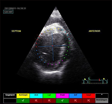
Fig. 1 Right parasternal short axis beneath the mitral valve: automatic placement of region of interest segments.
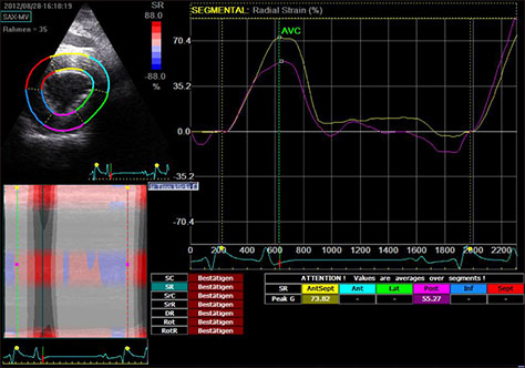
Fig. 2 Radial strain in the interventricular septum (purple) and the left ventricular free wall (yellow).

Fig. 3 Radial strain rate in the interventricular septum (purple) and the left ventricular free wall (yellow).
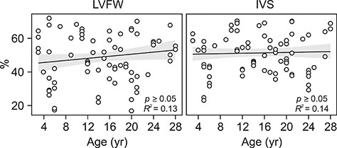
Fig. 4 Strain at different ages in the interventricular septum (IVS) and the left ventricular free wall (LVFW).
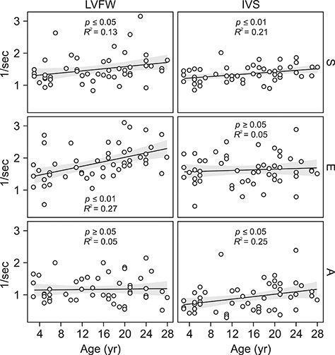
Fig. 5 Strain rate in the left ventricular free wall (LVFW) and the interventricular septum (IVS) at different ages. S, systolic maximum velocity; E, early diastolic filling; A, atrial release.
Reference
-
1. Amundsen BH, Helle-Valle T, Edvardsen T, Torp H, Crosby J, Lyseggen E, Støylen A, Ihlen H, Lima JA, Smiseth OA, Slørdahl SA. Noninvasive myocardial strain measurement by speckle tracking echocardiography: validation against sonomicrometry and tagged magnetic resonance imaging. J Am Coll Cardiol. 2006; 47:789–793.
Article2. Artis NJ, Oxborough DL, Williams G, Pepper CB, Tan LB. Two-dimensional strain imaging: a new echocardiographic advance with research and clinical applications. Int J Cardiol. 2008; 123:240–248.
Article3. Castro PL, Greenberg NL, Drinko J, Garcia MJ, Thomas JD. Potential pitfalls of strain rate imaging: angle dependency. Biomed Sci Instrum. 2000; 36:197–202.4. Cho GY, Chan J, Leano R, Strudwick M, Marwick TH. Comparison of two-dimensional speckle and tissue velocity based strain and validation with harmonic phase magnetic resonance imaging. Am J Cardiol. 2006; 97:1661–1666.
Article5. Dalen H, Thorstensen A, Aase SA, Ingul CB, Torp H, Vatten LJ, Stoylen A. Segmental and global longitudinal strain and strain rate based on echocardiography of 1266 healthy individuals: the HUNT study in Norway. Eur J Echocardiogr. 2010; 11:176–183.
Article6. Decloedt A, Verheyen T, Sys S, De Clercq D, van Loon G. Quantification of left ventricular longitudinal strain, strain rate, velocity, and displacement in healthy horses by 2-dimensional speckle tracking. J Vet Intern Med. 2011; 25:330–338.
Article7. Decloedt A, Verheyen T, Sys S, De Clercq D, van Loon G. Two-dimensional speckle tracking for quantification of left ventricular circumferential and radial wall motion in horses. Equine Vet J. 2013; 45:47–55.
Article8. D'hooge J, Heimdal A, Jamal F, Kukulski T, Bijnens B, Rademakers F, Hatle L, Suetens P, Sutherland GR. Regional strain and strain rate measurements by cardiac ultrasound: principles, implementation and limitations. Eur J Echocardiogr. 2000; 1:154–170.9. Di Salvo G, Russo MG, Paladini D, Pacileo G, Felicetti M, Ricci C, Cardaropoli D, Palma M, Caso P, Calabro R. Quantification of regional left and right ventricular longitudinal function in 75 normal fetuses using ultrasound-based strain rate and strain imaging. Ultrasound Med Biol. 2005; 31:1159–1162.
Article10. Dohi K, Suffoletto M, Murali S, Bazaz R, Gorcsan J. Benefit of cardiac resynchronization therapy to a patient with a narrow QRS complex and ventricular dyssynchrony identified by tissue synchronization imaging. Eur J Echocardiogr. 2005; 6:455–460.
Article11. Heimdal A, Støylen A, Torp H, Skjaerpe T. Real-time strain rate imaging of the left ventricle by ultrasound. J Am Soc Echocardiogr. 1998; 11:1013–1019.
Article12. Ihaka R. R: past and future history. Comput Sci Stat. 1998; 392–396.13. Jamal F, Strotmann J, Weidemann F, Kukulski T, D'hooge J, Bijnens B, Van de Werf F, De Scheerder I, Sutherland GR. Noninvasive quantification of the contractile reserve of stunned myocardium by ultrasonic strain rate and strain. Circulation. 2001; 104:1059–1065.
Article14. Kaluzynski K, Chen X, Emelianov SY, Skovoroda AR, O'Donnell M. Strain rate imaging using two-dimensional speckle tracking. IEEE Trans Ultrason Ferroelectr Freq Control. 2001; 48:1111–1123.
Article15. Kukulski T, Jamal F, Herbots L, D'hooge J, Bijnens B, Hatle L, De Scheerder I, Sutherland GR. Identification of acutely ischemic myocardium using ultrasonic strain measurements: a clinical study in patients undergoing coronary angioplasty. J Am Coll Cardiol. 2003; 41:810–819.
Article16. Kuznetsova T, Herbots L, Richart T, D'hooge J, Thijs L, Fagard RH, Herregods MC, Staessen JA. Left ventricular strain and strain rate in a general population. Eur Heart J. 2008; 29:2014–2023.
Article17. Lakatta EG. Cardiovascular aging research: the next horizons. J Am Geriatr Soc. 1999; 47:613–625.
Article18. Marcomichelakis J, Withers R, Newman GB, O'Brien K, Emanuel R. The relation of age to the thickness of the interventricular septum, the posterior left ventricular wall and their ratio. Int J Cardiol. 1983; 4:405–419.
Article19. Miyatake K, Yamagishi M, Tanaka N, Uematsu M, Yamazaki N, Mine Y, Sano A, Hirama M. New method for evaluating left ventricular wall motion by color-coded tissue Doppler imaging: in vitro and in vivo studies. J Am Coll Cardiol. 1995; 25:717–724.
Article20. Moore CC, Lugo-Olivieri CH, McVeigh ER, Zerhouni EA. Three-dimensional systolic strain patterns in the normal human left ventricle: characterization with tagged MR imaging. Radiology. 2000; 214:453–466.
Article21. Nagel D, Gehlen H. [The effectiveness of romifidine on myocardial function in horses with and without heart disease, evaluated with M-mode echocardiography and PW-tissue Doppler imaging]. Berl Munch Tierarztl Wochenschr. 2013; 126:436–443. German.22. Olivetti G, Melissari M, Capasso JM, Anversa P. Cardiomyopathy of the aging human heart. Myocyte loss and reactive cellular hypertrophy. Circ Res. 1991; 68:1560–1568.
Article23. Sage AM. Cardiac disease in the geriatric horse. Vet Clin North Am Equine Pract. 2002; 18:575–589. viii
Article24. Schefer KD, Bitschnau C, Weishaupt MA, Schwarzwald CC. Quantitative analysis of stress echocardiograms in healthy horses with 2-dimensional (2D) echocardiography, anatomical M-mode, tissue Doppler imaging, and 2D speckle tracking. J Vet Intern Med. 2010; 24:918–931.
Article25. Schwarzwald CC, Schober KE, Berli AS, Bonagura JD. Left ventricular radial and circumferential wall motion analysis in horses using strain, strain rate, and displacement by 2D speckle tracking. J Vet Intern Med. 2009; 23:890–900.
Article26. Schwarzwald CC, Schober KE, Bonagura JD. Methods and reliability of tissue Doppler imaging for assessment of left ventricular radial wall motion in horses. J Vet Intern Med. 2009; 23:643–652.
Article27. Sengupta PP, Krishnamoorthy VK, Korinek J, Narula J, Vannan MA, Lester SJ, Tajik JA, Seward JB, Khandheria BK, Belohlavek M. Left ventricular form and function revisited: applied translational science to cardiovascular ultrasound imaging. J Am Soc Echocardiogr. 2007; 20:539–551.
Article28. Stadler P, Robine F. [Die Kardiometrie beim gesunden Warmblutpferd mit Hilfe der Schnittbildechographie im B-Mode]. Pferdeheilkunde. 1996; 12:35–43. German.29. Stoylen A, Heimdal A, Bjornstad K, Torp HG, Skjaerpe T. Strain rate imaging by ultrasound in the diagnosis of regional dysfunction of the left ventricle. Echocardiography. 1999; 16:321–329.
Article30. Sun JP, Chinchoy E, Donal E, Popović ZB, Perlic G, Asher CR, Greenberg NL, Grimm RA, Wilkoff BL, Thomas JD. Evaluation of ventricular synchrony using novel Doppler echocardiographic indices in patients with heart failure receiving cardiac resynchronization therapy. J Am Soc Echocardiogr. 2004; 17:845–850.
Article31. Sun JP, Popović ZB, Greenberg NL, Xu XF, Asher CR, Stewart WJ, Thomas JD. Noninvasive quantification of regional myocardial function using Doppler-derived velocity, displacement, strain rate, and strain in healthy volunteers: effects of aging. J Am Soc Echocardiogr. 2004; 17:132–138.
Article32. Teske AJ, De Boeck BW, Melman PG, Sieswerda GT, Doevendans PA, Cramer MJ. Echocardiographic quantification of myocardial function using tissue deformation imaging, a guide to image acquisition and analysis using tissue Doppler and speckle tracking. Cardiovasc Ultrasound. 2007; 5:27.
Article33. Thomas JD, Popović ZB. Assessment of left ventricular function by cardiac ultrasound. J Am Coll Cardiol. 2006; 48:2012–2025.
Article34. Voigt I, Mansi T, Ionasec RI, Mengue EA, Houle H, Georgescu B, Hornegger J, Comaniciu D. Robust physically-constrained modeling of the mitral valve and subvalvular apparatus. In : 14th International Conference on Medical Image Computing & Computer Assisted Intervention: MICCAI 2011; 18–22 Sep 2011; Toronto, Canada.35. Wei JY. Age and the cardiovascular system. N Engl J Med. 1992; 327:1735–1739.
Article
- Full Text Links
-
- Actions
-
Cited
- CITED
-
- Close
- Share
-
- Similar articles
-
- Speckle tracking ultrasonography as a new tool to assess diaphragmatic function: a feasibility study
- Current Status of 3-Dimensional Speckle Tracking Echocardiography: A Review from Our Experiences
- Endurance and Strength Athlete's Heart: Analysis of Myocardial Deformation by Speckle Tracking Echocardiography
- Preliminary study on the effects of pergolide on left ventricular function in the horses with pituitary pars intermedia dysfunction
- Two-dimensional speckle-tracking of antral contraction in dogs
