Global Circumferential Strain by 2-Dimensional Speckle Tracking Method for the Evaluation of the Left Ventricular Function
- Affiliations
-
- 1Division of Cardiology, Department of Internal Medicine, Gachon University of Medicine and Science, Gil Medical Center, Incheon, Korea. msshin@gilhospital.com
- 2Division of Nephrology, Department of Internal Medicine, Gachon University of Medicine and Science, Gil Medical Center, Incheon, Korea.
- KMID: 2225772
- DOI: http://doi.org/10.4070/kcj.2008.38.7.379
Abstract
-
BACKGROUND AND OBJECTIVES: The speckle tracking method using 2-dimensional (2D) echocardiography is not affected by the tethering of neighboring segments and angulation. Global circumferential strain (GCS) of the left ventricle (LV) has been suggested as a systolic index and correlated with LV contractility. The purpose of this study was to investigate whether acute changes in preload affect global circumferential strain and to evaluate the usefulness of GCS by the speckle tracking method.
SUBJECTS AND METHODS
2D echocardiography was performed in 69 patients with end-stage renal disease before and after hemodialysis to measure the LV end-diastolic volume and LV ejection fraction. 2D images were acquired from the short-axis view of the mid-LV for the evaluation of GCS.
RESULTS
Mean LV end-diastolic volume significantly decreased from 91.2+/-33.3 mL to 72.3+/-32.0 mL (p+/-0.05), and LV ejection fraction decreased from 63.6+/-13.1% to 60.0+/-11.2% (p=0.006) after hemodialysis. However, mean GCS showed no significant change after hemodialysis (17.2+/-5.3% vs. 16.6+/-4.7%, p=0.13). GCS was found to be well correlated with LV ejection fraction (r=0.54, p<0.05) and peak systolic mitral annular velocity (r=0.46, p=0.000), but not with LV preload (r=0.06, p=0.622).
CONCLUSION
GCS using the speckle tracking method is a useful index for the evaluation of LV systolic function because it is not affected by acute preload change and is correlated with LV ejection fraction and peak systolic mitral annular velocity.
MeSH Terms
Figure
-
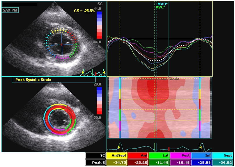
Fig. 1 Speckle tracking method using a 2D echocardiographic image. Automatic frame-by-frame tracking of natural acoustic markers around the traced line was used to measure the global circumferential strain along the selected region of interest at the level of the mid-LV.
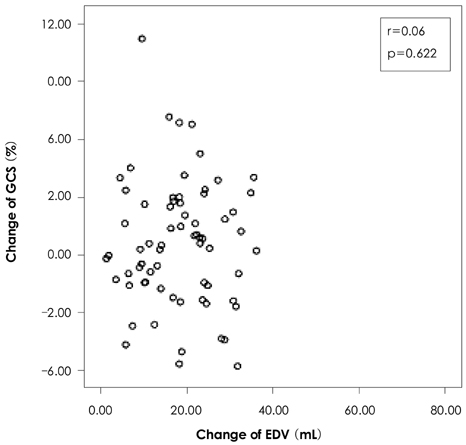
Fig. 2 Correlation between left ventricular preload (end-diastolic volume, EDV) change and global circumferential strain (GCS) change.
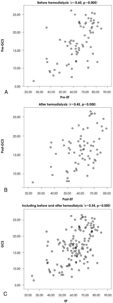
Fig. 3 Correlation between ejection fraction (EF) and global circumferential strain (GCS). A: pre-hemodialysis. B: post-hemodialysis. C: including pre- and post-hemodialysis. Pre-GCS: GCS before hemodialysis, pre-EF: EF before hemodialysis, post-GCS: GCS after hemodialysis, post-EF: EF after hemodialysis.
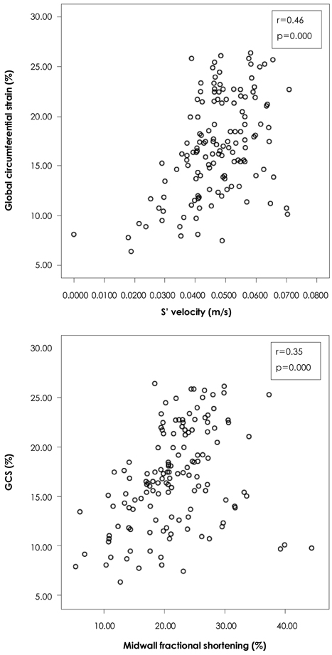
Fig. 4 Correlation between global circumferential strain (GCS) and mitral annular peak systolic velocity (S') (upper graph) and midwall fractional shortening (lower graph).

Fig. 5 Receiver operating characteristic (ROC) curve of global circumferential strain (GCS) (area under the curve, AUC 0.765). Sensitivity was 72.7% and specificity was 54.3% when a cut-off GCS value of 17.11% was used to detect normal left ventricular ejection fraction.
Reference
-
1. Sutherland GR, Stewart MJ, Groundstroem KW, et al. Color Doppler myocardial imaging: a new technique for the assessment of myocardial function. J Am Soc Echocardiogr. 1994. 7:441–458.2. Pislaru C, Abraham TP, Belohlavek M. Strain and strain rate echocardiography. Curr Opin Cardiol. 2002. 17:443–454.3. Sutherland GR, Di Salvo G, Claus P, D'hooge J, Bijnens B. Strain and strain rate imaging: a new clinical approach to quantifying regional myocardial function. J Am Soc Echocardiogr. 2004. 17:788–802.4. Tei C. New non-invasive index for combined systolic and diastolic ventricular function. J Cardiol. 1995. 26:135–136.5. Agricola E, Galderisi M, Oppizzi M, et al. Pulsed tissue Doppler imaging detects early myocardial dysfunction in asymptomatic patients with severe mitral regurgitation. Heart. 2004. 90:406–410.6. Barberato SH, Pecoits Filho R. Influence of preload reduction on Tei index and other Doppler echocardiographic parameters of left ventricular function. Arq Bras Cardiol. 2006. 86:425–431.7. Abraham TP, Nishimura RA, Holmes DR Jr, Belohlavek M, Seward JB. Strain rate imaging for assessment of regional myocardial function: results from a clinical model of septal ablation. Circulation. 2002. 105:1403–1406.8. Greenberg NL, Firstenberg MS, Castro PL, et al. Doppler-derived myocardial systolic strain rate is a strong index of left ventricular contractility. Circulation. 2002. 105:99–105.9. Serri K, Reant P, Lafitte M, et al. Global and regional myocardial function quantification by two-dimensional strain: application in hypertrophic cardiomyopathy. J Am Coll Cardiol. 2006. 47:1175–1181.10. Marwick TH. Measurement of strain and strain rate by echocardiography. J Am Coll Cardiol. 2006. 47:1313–1327.11. Leitman M, Lysyansky P, Sidenko S, et al. Two-dimensional strain-A novel software for real-time quantitative echocardiographic assessment of myocardial function. J Am Soc Echocardiogr. 2004. 17:1021–1029.12. Modesto KM, Cauduro S, Dispenzieri A, et al. Two-dimensional acoustic pattern derived strain parameters closely correlate with one-dimensional tissue Doppler derived strain measurements. Eur J Echocardiogr. 2006. 7:315–321.13. Park KH, Song JK, Suh IW, et al. The usefulness of 2-dimensional longitudinal strain for prediction of the postoperative left ventricular systolic function in patients with valvular heart disease causing volume overloading. Korean Circ J. 2006. 36:272–278.14. Reisner SA, Lysyansky P, Aqmon Y, Mutlak D, Lessick J, Friedman Z. Global longitudinal strain: a novel index of left ventricular systolic function. J Am Soc Echocardiogr. 2004. 17:630–633.15. Helm RH, Leclercq C, Faris OP, et al. Cardiac dyssynchrony analysis using circumferential versus longitudinal strain. Circulation. 2005. 111:2760–2767.16. Chakko S, Girgis I, Contreras G, Perez G, Kessler KM, Myerburg RJ. Effects of hemodialysis on left ventricular diastolic filling. Am J Cardiol. 1997. 79:106–108.17. Vancheri F, Barberi O, Cammalleri G, et al. Echopolycardiographic evaluation of left ventricular function after hemodialysis. G Ital Cardiol. 1985. 15:673–676.18. Nixon JV, Mitchell JH, McPhaul JJ Jr, Henrich WL. Effect of hemodialysis on left ventricular function: dissociation of changes in filling volume and in contractile state. J Clin Invest. 1983. 71:377–384.19. Devereaux RB, Alonso DR, Lutas EM, et al. Echocardiographic assessment of left ventricular hypertrophy: comparison to necropsy findings. Am J Cardiol. 1986. 57:450–458.20. de Simone G, Devereux RB, Roman MJ, et al. Assessment of left ventricular function by the midwall fractional shortening/end-systolic stress relation in human hypertension. J Am Coll Cardiol. 1994. 23:1444–1451.21. Jung HO. Evaluation of midwall function using echocardiography. J Cardiovasc Ultrasound. 2007. 15:115–120.22. Gilman G, Khandheria BK, Hagen ME, Abraham TP, Seward JB, Belohlavek M. Strain rate and strain: a step-by-step approach to image and data acquisition. J Am Soc Echocardiogr. 2004. 17:1011–1020.23. D'hooge J, Heimdal A, Jamal F, et al. Regional strain and strain rate measurements by cardiac ultrasound: principles, implementation and limitations. Eur J Echocardiogr. 2000. 1:154–170.24. Amundsen BH, Helle-Valle T, Edvardsen T, et al. Noninvasive myocardial strain measurement by speckle tracking echocardiography: validation against sonomicrometry and tagged magnetic resonance imaging. J Am Coll Cardiol. 2006. 47:789–793.25. Anderson NH, Terkelsen CJ, Sloth E, Poulsen SH. Influence of preload alterations on parameters of systolic left ventricular long-axis function: a Doppler tissue study. J Am Soc Echocardiogr. 2004. 17:941–947.26. Eidem BW, McMahon CJ, Ayres NA, et al. Impact of chronic left ventricular preload and afterload on Doppler tissue imaging velocities: a study in congenital heart disease. J Am Soc Echocardiogr. 2005. 18:830–838.27. Dalsgaard M, Snyder EM, Kjaergaard J, Johnson BD, Hassager C, Oh JK. Isovolumic acceleration measured by tissue Doppler echocardiography is preload independent in healthy subjects. Echocardiography. 2007. 24:572–579.28. Oğuzhan A, Arinç H, Abaci A, et al. Preload dependence of Doppler tissue imaging derived indexes of left ventricular diastolic function. Echocardiography. 2005. 22:320–325.29. Hung KC, Huang HL, Chu CM, Yeh KH, Fang JT, Lin FC. Effects of altered volume loading on left ventricular hemodynamics and diastolic filling during hemodialysis. Ren Fail. 2004. 26:141–147.30. Hung KC, Huang HL, Chu CM, et al. Evaluating preload dependence of a novel Doppler application in assessment of left ventricular diastolic function during hemodialysis. Am J Kidney Dis. 2004. 43:1040–1046.31. Amà R, Segers P, Roosens C, Claessens T, Verdonck P, Poelaert J. The effects of load on systolic mitral annular velocity by tissue Doppler imaging. Anesth Analg. 2004. 99:332–338.
- Full Text Links
-
- Actions
-
Cited
- CITED
-
- Close
- Share
-
- Similar articles
-
- Alterations in Left Ventricular Circumferential Motion in Children with Dilated Cardiomyopathy
- Left Ventricular Radial Strain in Children with Dilated Cardiomyopathy: Analyzed with Two Dimensional Speckle Tracking Imaging Method
- Utility of Global Strain by Two-Dimensional and Three-Dimensional Speckle Tracking for Assessing Left Ventricular Diastolic Function: Comparison with Pressure Wire Analysis
- Current Status of 3-Dimensional Speckle Tracking Echocardiography: A Review from Our Experiences
- Two-dimensional speckle-tracking of antral contraction in dogs
