Identification of Dermatophytes Using Multiplex Polymerase Chain Reaction
- Affiliations
-
- 1Department of Dermatology, Konkuk University School of Medicine, Seoul, Korea. 20050078@kuh.ac.kr
- KMID: 2171921
- DOI: http://doi.org/10.5021/ad.2011.23.3.304
Abstract
- BACKGROUND
Multiplex polymerase chain reaction (PCR) allows more than two target DNA molecules to be amplified with more than two primers. This method is also useful for detecting various other organisms simultaneously within a single test tube, and the scope of its use has been expanding widely in the field of clinical microbiology in recent years.
OBJECTIVE
To assess the value of multiplex PCR in identification of dermatophytes.
METHODS
Using three specially-designed primers which contained the ITS1-2, 18S rRNA, and 28S rRNA regions, three cycles of PCR were performed on 11 standard strains and scales were collected from 73 patients with fungal infection.
RESULTS
The 11 standard strains were successfully identified with analysis of band patterns of ITS1-2, 18S rRNA, and 28S rRNA, obtained from PCR. Based on this information, the causative organisms in 73 patients with fungal infection were revealed to be T. rubrum in 69 cases, T. menta in 1 case, T. tonsurans in 2 cases, and M. gypseum in one case.
CONCLUSION
With three cycles of PCR using three sets of primers, 11 standard strains and the clinical strains from 73 patients with fungal infection were successfully identified.
Keyword
MeSH Terms
Figure
-

Fig. 1 PCR amplification from dermatophyte standard strains with (A) the ITS1-2 region, (B) the 18S ribosomal RNA region and (C) the 28S ribosomal RNA region. Lanes: M, molecular marker; 1. Epidermophyton floccosum (CBS 358.93); 2. Microsporum canis (IFM 45829); 3. Microsporum audouinii (IFM 5294 [CBS 319.31]); 4. Trichophyton violaceum (CBS 319.31); 5. Trichophyton rubrum (KCTC 6375(ATCC 28188)); 6. Trichophyton mentagrophytes var. mentagrophytes (CBS 126.34); 7. Trichophyton tonsurans (CBS 483.76); 8. Trichophyton mentagrophytes var. interdigitale (IFM 53931); 9. Trichophyton verrucosum (CBS 134.66); 10. Microsporum gypseum (IFM 5292); 11. Microsporum fulvum (IFM 5318); 12. Control (dH2O). PCR: polymerase chain reaction.
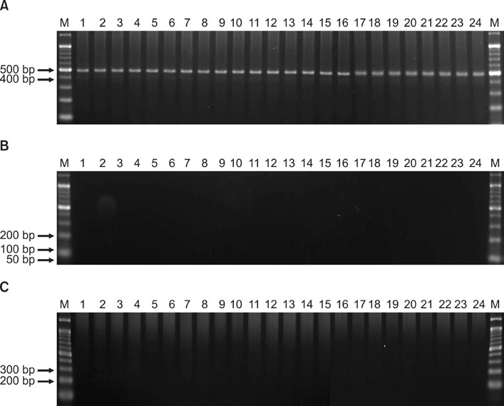
Fig. 2 PCR amplification of (A) the ITS1-2 region, (B) the 18S ribosomal RNA region and (C) the 28S ribosomal RNA region from onychomycosis. Lanes: M, molecular marker; 1~24. Trichophyton rubrum. PCR: polymerase chain reaction.
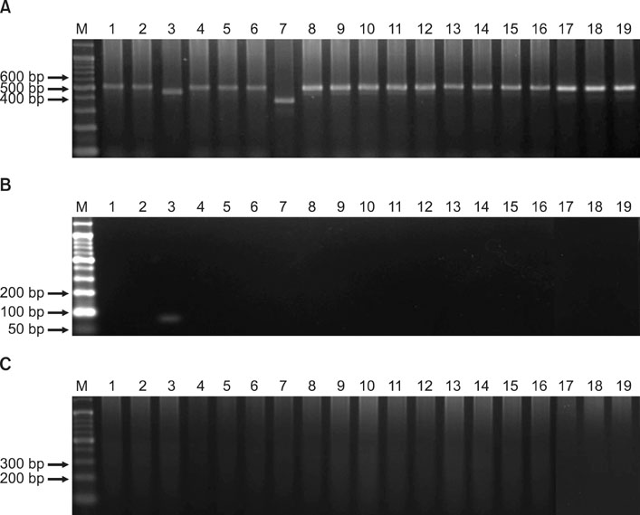
Fig. 3 PCR amplification of (A) the ITS1-2 region, (B) the 18S ribosomal RNA region and (C) the 28S ribosomal RNA region from tinea pedis. Lanes: M, molecular marker; 3. Trichophyton mentagrophytes var. mentagrophytes, 7. Trichophyton tonsurans. The other lanes were Trichophyton rubrum. PCR: polymerase chain reaction.
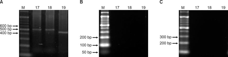
Fig. 4 PCR amplification of (A) the ITS1-2 region, (B) the 18S ribosomal RNA region and (C) the 28S ribosomal RNA region from tinea capitis. Lanes: M, molecular marker; 17~18. Trichophyton rubrum, 19. Trichophyton tonsurans. PCR: polymerase chain reaction.

Fig. 5 PCR amplification of (A) the ITS1-2 region, (B) the 18S ribosomal RNA region and (C) the 28S ribosomal RNA region from tinea corporis. Lanes: M, molecular marker; 7. Microsporum gypseum. The other lanes were Trichophyton rubrum. PCR: polymerase chain reaction.
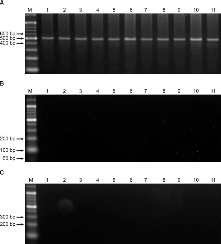
Fig. 6 PCR amplification of (A) the ITS1-2 region, (B) the 18S ribosomal RNA region and (C) the 28S ribosomal RNA region from tinea cruris. Lanes: M, molecular marker; 1~11. Trichophyton rubrum. PCR: polymerase chain reaction.
Reference
-
1. Verma S, Heffernan MP. Wolff K, Goldsmith LA, Katz SI, Gilchrest BA, Paller AS, Leffell DJ, editors. Superficial fungal infection: dermatophytosis, onychomycosis, tinea nigra, piedra. Fitzpatrick's dermatology in general medicine. 2008. 7th ed. New York: McGraw-Hill;1817–1821.2. Gupta AK, Tu LQ. Dermatophytes: diagnosis and treatment. J Am Acad Dermatol. 2006. 54:1050–1055.
Article3. Lee KJ, Kim JE, Park HJ, Lee JY, Cho BK. A case of Trychophyton mentagrophytes var. erinacei infection from a patient's pet hedgehog. Korean J Med Mycol. 2009. 14:98–102.4. Jung HD, Chi SG, Lee WJ, Jun JB, Suh SB, Bang YJ, et al. A case of tinea corporis caused by Microsporum ferrugineum. Korean J Med Mycol. 2008. 13:37–40.5. Jun JB, Kim YD. The epidemiological, clinical and mycological studies on trichophytosis gladiatorum prevailing among Korean wrestlers. Korean J Med Mycol. 2004. 9:28–44.6. Kim KH. Identification of dermatophytes. Korean J Med Mycol. 1997. 2:1–8.7. Lee YW. Molecular analysis for identification and classification of dermatophytes. Korean J Dermatol. 2010. 48:Suppl. 1. 82.8. Summerbell RC, Kane J. Kane J, Summerbell RC, Sigler L, Krajden S, Land G, editors. Physiologic and other special tests for identifying dermatophytes. Laboratory handbook of dermatophytes. 1997. Belmont: Star Publishing Co.;45–77.9. Yang G, Zhang M, Li W, An L. Direct species identification of common pathogenic dermatophyte fungi in clinical specimens by semi-nested PCR and restriction fragment length polymorphism. Mycopathologia. 2008. 166:203–208.
Article10. Garg J, Tilak R, Singh S, Gulati AK, Garg A, Prakash P, et al. Evaluation of pan-dermatophyte nested PCR in diagnosis of onychomycosis. J Clin Microbiol. 2007. 45:3443–3445.
Article11. Kim JA, Moon SE, Kwon TE, Yu HJ, Cho BK, Lee KH, et al. Identification of dermatophytes by mycological tests and random amplified polymorphic DNA analysis. Korean J Dermatol. 2001. 39:168–175.12. Kim JY, Hwang YJ, Ko JH, Oh BH, Lee YW, Choe YB, et al. A case of kerion celsi caused by Trichophyton verrucosum. Korean J Med Mycol. 2010. 15:83–87.13. Lim SW, Shin MG, Lim JY, Yun SJ, Kim SJ, Lee SC, et al. Nested PCR for detection of Malassezia species from patient skin scales and clinical strains. Korean J Dermatol. 2008. 46:446–452.14. Lee MK, Kim HR, Lee YJ. Identification of Candida species by multiplex polymerase chain reaction. Korean J Clin Microbiol. 2006. 9:119–124.15. Suh MK, Kim BC, Kim JC. Phylogeny and taxonomy of the dermatophytes using sequence analysis of the ribosomal internal transcribed spacer 1 region. Korean J Dermatol. 2000. 38:1186–1193.16. Seebacher C, Bouchara JP, Mignon B. Updates on the epidemiology of dermatophyte infections. Mycopathologia. 2008. 166:335–352.
Article17. Dragos V, Lunder M. Lack of efficacy of 6-week treatment with oral terbinafine for tinea capitis due to Microsporum canis in children. Pediatr Dermatol. 1997. 14:46–48.
Article18. Fleece D, Gaughan JP, Aronoff SC. Griseofulvin versus terbinafine in the treatment of tinea capitis: a meta-analysis of randomized, clinical trials. Pediatrics. 2004. 114:1312–1315.
Article
- Full Text Links
-
- Actions
-
Cited
- CITED
-
- Close
- Share
-
- Similar articles
-
- Rapid Diagnosis of Duchenne Muscular Dystrophy DMD by Multiplex Polymerase Chain Reaction PCR using Uncultured Amniocytes
- Identification of Candida Species by Multiplex Polymerase Chain Reaction
- Multiplex PCR Assay for Identification of Mycobacterial Species Isolated from Liquid Cultures
- Identification of Mycobacterium Species by Multiplex PCR-Restriction Fragment Length Polymorphism Assay
- Multiplex Real-time Polymerase Chain Reaction Assays for Simultaneous Detection of
Vibrio cholerae ,Vibrio parahaemolyticus , andVibrio vulnificus
