Enhanced Anti-tumor Reactivity of Cytotoxic T Lymphocytes Expressing PD-1 Decoy
- Affiliations
-
- 1Research Institute National Cancer Center, Goyang, Gyeonggi-do 10408, Korea.
- 2Department of Biochemistry and Molecular Biology and Department of Biomedical Sciences, Seoul National University College of Medicine, Seoul 03080, Korea. khchoi@snu.ac.kr
- KMID: 2168054
- DOI: http://doi.org/10.4110/in.2016.16.2.134
Abstract
- Programmed death-1 (PD-1) is a strong negative regulator of T lymphocytes in tumor-microenvironment. By engaging PD-1 ligand (PD-L1) on tumor cells, PD-1 on T cell surface inhibits anti-tumor reactivity of tumor-infiltrating T cells. Systemic blockade of PD-1 function using blocking antibodies has shown significant therapeutic efficacy in clinical trials. However, approximately 10 to 15% of treated patients exhibited serious autoimmune responses due to the activation of self-reactive lymphocytes. To achieve selective activation of tumor-specific T cells, we generated T cells expressing a dominant-negative deletion mutant of PD-1 (PD-1 decoy) via retroviral transduction. PD-1 decoy increased IFN-γ secretion of antigen-specific T cells in response to tumor cells expressing the cognate antigen. Adoptive transfer of PD-1 decoy-expressing T cells into tumor-bearing mice potentiated T cell-mediated tumor regression. Thus, T cell-specific blockade of PD-1 could be a useful strategy for enhancing both efficacy and safety of anti-tumor T cell therapy.
Keyword
MeSH Terms
Figure
-
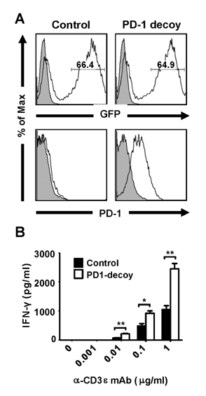
Figure 1 Overexpression of PD-1 decoy increases IFN-γ secretion from T cells. Activated B6 splenic T cells were transduced with retrovirus carrying either a control vector (pMIG-w) or PD-1 decoy and rested for 3 days in the absence of stimulation. (A) Retroviral transduction efficiency was measured by flow cytometry using GFP. The GFP positive populations were gated and the expression levels of PD-1 were analyzed. PD-1 expression in the control represents the levels of endogenous PD-1, while the PD-1 decoy sample shows the levels of both endogenous and the decoy receptor. (Filled gray area: Isotype control, Percentage of GFP positive cells indicated inside histograms) (B) GFP positive T cells were sorted and stimulated with anti-CD3 in the presence of irradiated splenocytes for 48 hours. IFN-γ in the cultured supernatants was measured by ELISA (Student's t-test, *p<0.05, **p<0.01). Results are representative of 3 independent experiments.

Figure 2 Enhanced anti-tumor reactivity of OT-I T cells carrying PD-1 decoy. OVA-specific CD8 T cells (OT-I cells) were transduced with retroviruses carrying control vector or PD-1 decoy as described in the methods and materials. (A) Transduction efficiency and PD-1 decoy expression of OT-I cells were measured as described in Fig. 1A. (B) Expression of PD-L1 on OVA-expressing tumor cell lines was analyzed by flow cytometry. (A-B, Gray filled area: Isotype control, Percentage of GFP positive cells indicated inside histograms) (C) The retrovirus-transduced OT-I cells were co-cultured with OVA-expressing tumor cell lines for 24 hours, and IFN-γ in the cultured supernatants was measured by ELISA (Student's t-test, ns; not significant, *p<0.05, **p<0.01, ****p<0.0001). The transduced OT-I cells were rested for 3 days in the absence of stimulation and sorted by GFP expression before co-culture with the tumor cells. Results are representative of 2~3 independent experiments (A-C).
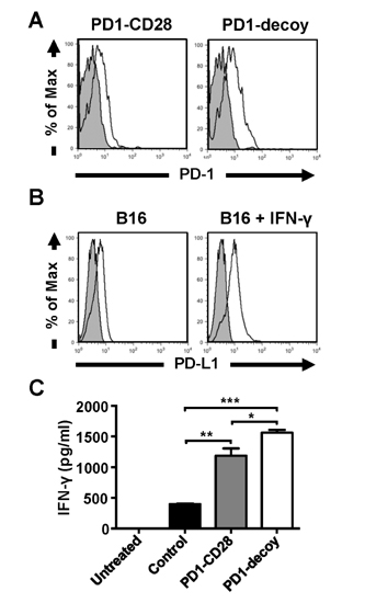
Figure 3 The PD-1-CD28 chimera does not improve the anti-tumor reactivity of T lymphocytes compared to PD-1 decoy. B16 melanoma antigen (gp100)-specific CD8 T cells (Pmel-1 cells) were transduced with retroviruses carrying PD-1-CD28 chimera or PD-1 decoy. (A) Expression levels of PD-1-CD28 chimera and PD-1 decoy in GFP-positive Pmel-1 populations were analyzed by flow cytometry. (B) B16 melanoma cells were either left untreated or treated with IFN-γ (20 ng/ml) for 48 hours. Then, PD-L1 expression was determined by flow cytometry. (C) Retrovirus-transduced GFP positive Pmel-1 cells were sorted by flow cytometry. The sorted cells (1×10⁵) were co-cultured with IFN-γ-treated B16 melanoma cells (1×10⁴) for 48 hours. IFN-γ in the cultured supernatants was quantified by ELISA (Student's t-test, *p<0.05, **p<0.01, ***p<0.001).

Figure 4 PD-1 decoy potentiates anti-tumor therapeutic efficacy of adoptively transferred T cells. B6 mice were injected with OVA-expressing E.G7 cells (2×10⁶) subcutaneously. After 7 days, the mice were either left untreated or intravenously injected with control vector (pMIG-w)-transduced (2×10 6) or PD-1 decoy-transduced (2×10⁶) OT-I cells. The retrovirus-transduced OT-I cells were rested for 3 days before adoptive transfer. The mean tumor size of 10 mice per group was recorded (*p=0.0039, Wilcoxon matched-pairs test). Results are representative of 2 independent experiments.
Reference
-
1. Kyi C, Postow MA. Checkpoint blocking antibodies in cancer immunotherapy. FEBS Lett. 2014; 588:368–376.
Article2. Henick BS, Herbst RS, Goldberg SB. The PD-1 pathway as a therapeutic target to overcome immune escape mechanisms in cancer. Expert Opin Ther Targets. 2014; 18:1407–1420.
Article3. Topalian SL, Hodi FS, Brahmer JR, Gettinger SN, Smith DC, McDermott DF, Powderly JD, Carvajal RD, Sosman JA, Atkins MB, Leming PD, Spigel DR, Antonia SJ, Horn L, Drake CG, Pardoll DM, Chen L, Sharfman WH, Anders RA, Taube JM, McMiller TL, Xu H, Korman AJ, Jure-Kunkel M, Agrawal S, McDonald D, Kollia GD, Gupta A, Wigginton JM, Sznol M. Safety, activity, and immune correlates of anti-PD-1 antibody in cancer. N Engl J Med. 2012; 366:2443–2454.
Article4. Topalian SL, Sznol M, McDermott DF, Kluger HM, Carvajal RD, Sharfman WH, Brahmer JR, Lawrence DP, Atkins MB, Powderly JD, Leming PD, Lipson EJ, Puzanov I, Smith DC, Taube JM, Wigginton JM, Kollia GD, Gupta A, Pardoll DM, Sosman JA, Hodi FS. Survival, durable tumor remission, and long-term safety in patients with advanced melanoma receiving nivolumab. J Clin Oncol. 2014; 32:1020–1030.
Article5. Hamid O, Robert C, Daud A, Hodi FS, Hwu WJ, Kefford R, Wolchok JD, Hersey P, Joseph RW, Weber JS, Dronca R, Gangadhar TC, Patnaik A, Zarour H, Joshua AM, Gergich K, Elassaiss-Schaap J, Algazi A, Mateus C, Boasberg P, Tumeh PC, Chmielowski B, Ebbinghaus SW, Li XN, Kang SP, Ribas A. Safety and tumor responses with lambrolizumab (anti-PD-1) in melanoma. N Engl J Med. 2013; 369:134–144.
Article6. Philips GK, Atkins M. Therapeutic uses of anti-PD-1 and anti-PD-L1 antibodies. Int Immunol. 2015; 27:39–46.
Article7. Okazaki T, Honjo T. PD-1 and PD-1 ligands: from discovery to clinical application. Int Immunol. 2007; 19:813–824.
Article8. Agata Y, Kawasaki A, Nishimura H, Ishida Y, Tsubata T, Yagita H, Honjo T. Expression of the PD-1 antigen on the surface of stimulated mouse T and B lymphocytes. Int Immunol. 1996; 8:765–772.
Article9. Dong H, Zhu G, Tamada K, Chen L. B7-H1, a third member of the B7 family, co-stimulates T-cell proliferation and interleukin-10 secretion. Nat Med. 1999; 5:1365–1369.
Article10. Latchman Y, Wood CR, Chernova T, Chaudhary D, Borde M, Chernova I, Iwai Y, Long AJ, Brown JA, Nunes R, Greenfield EA, Bourque K, Boussiotis VA, Carter LL, Carreno BM, Malenkovich N, Nishimura H, Okazaki T, Honjo T, Sharpe AH, Freeman GJ. PD-L2 is a second ligand for PD-1 and inhibits T cell activation. Nat Immunol. 2001; 2:261–268.
Article11. Ishida M, Iwai Y, Tanaka Y, Okazaki T, Freeman GJ, Minato N, Honjo T. Differential expression of PD-L1 and PD-L2, ligands for an inhibitory receptor PD-1, in the cells of lymphohematopoietic tissues. Immunol Lett. 2002; 84:57–62.
Article12. Liang SC, Latchman YE, Buhlmann JE, Tomczak MF, Horwitz BH, Freeman GJ, Sharpe AH. Regulation of PD-1, PD-L1, and PD-L2 expression during normal and autoimmune responses. Eur J Immunol. 2003; 33:2706–2716.
Article13. Dong H, Strome SE, Salomao DR, Tamura H, Hirano F, Flies DB, Roche PC, Lu J, Zhu G, Tamada K, Lennon VA, Celis E, Chen L. Tumor-associated B7-H1 promotes T-cell apoptosis: a potential mechanism of immune evasion. Nat Med. 2002; 8:793–800.
Article14. Chemnitz JM, Parry RV, Nichols KE, June CH, Riley JL. SHP-1 and SHP-2 associate with immunoreceptor tyrosine-based switch motif of programmed death 1 upon primary human T cell stimulation, but only receptor ligation prevents T cell activation. J Immunol. 2004; 173:945–954.
Article15. Okazaki T, Maeda A, Nishimura H, Kurosaki T, Honjo T. PD-1 immunoreceptor inhibits B cell receptor-mediated signaling by recruiting src homology 2-domain-containing tyrosine phosphatase 2 to phosphotyrosine. Proc Natl Acad Sci U S A. 2001; 98:13866–13871.
Article16. Nishimura H, Nose M, Hiai H, Minato N, Honjo T. Development of lupus-like autoimmune diseases by disruption of the PD-1 gene encoding an ITIM motif-carrying immunoreceptor. Immunity. 1999; 11:141–151.
Article17. Nishimura H, Okazaki T, Tanaka Y, Nakatani K, Hara M, Matsumori A, Sasayama S, Mizoguchi A, Hiai H, Minato N, Honjo T. Autoimmune dilated cardiomyopathy in PD-1 receptor-deficient mice. Science. 2001; 291:319–322.
Article18. Muenst S, Soysal SD, Tzankov A, Hoeller S. The PD-1/PD-L1 pathway: biological background and clinical relevance of an emerging treatment target in immunotherapy. Expert Opin Ther Targets. 2015; 19:201–211.
Article19. Naidoo J, Page DB, Li BT, Connell LC, Schindler K, Lacouture ME, Postow MA, Wolchok JD. Toxicities of the anti-PD-1 and anti-PD-L1 immune checkpoint antibodies. Ann Oncol. 2015; 26:2375–2391.
Article20. Weber JS, Kahler KC, Hauschild A. Management of immune-related adverse events and kinetics of response with ipilimumab. J Clin Oncol. 2012; 30:2691–2697.
Article21. Shin JH, Park HB, Oh YM, Lim DP, Lee JE, Seo HH, Lee SJ, Eom HS, Kim IH, Lee SH, Choi K. Positive conversion of negative signaling of CTLA4 potentiates antitumor efficacy of adoptive T-cell therapy in murine tumor models. Blood. 2012; 119:5678–5687.
Article22. Prosser ME, Brown CE, Shami AF, Forman SJ, Jensen MC. Tumor PD-L1 co-stimulates primary human CD8(+) cytotoxic T cells modified to express a PD1:CD28 chimeric receptor. Mol Immunol. 2012; 51:263–272.
Article
- Full Text Links
-
- Actions
-
Cited
- CITED
-
- Close
- Share
-
- Similar articles
-
- Cytolytic activity of mitogen activated old and young mouse spleen cells against tumor target cells expressing high or low levels of Fas antigen
- Targeting TM4SF1 promotes tumor senescence enhancing CD8+ T cell cytotoxic function in hepatocellular carcinoma
- IL-17-Producing Cells in Tumor Immunity: Friends or Foes?
- Two-Round Mixed Lymphocyte Reaction for Evaluation of the Functional Activities of Anti-PD-1 and Immunomodulators
- Immunohistochemical expression of programmed death-ligand 1 and CD8 in glioblastomas
