Imaging Sci Dent.
2013 Sep;43(3):191-196. 10.5624/isd.2013.43.3.191.
Evaluation of the accuracy of linear and angular measurements on panoramic radiographs taken at different positions
- Affiliations
-
- 1Department of Oral and Maxillofacial Radiology, Dental School, Shahid Beheshti University of Medical Sciences, Tehran, Iran. emadi_ne@yahoo.com
- 2Department of Oral and Maxillofacial Radiology, Dental School, Ilam University of Medical Sciences, Ilam, Iran.
- KMID: 2167457
- DOI: http://doi.org/10.5624/isd.2013.43.3.191
Abstract
- PURPOSE
This study assessed the accuracy of linear and angular measurements on panoramic radiographs taken at different positions in vitro.
MATERIALS AND METHODS
Two acrylic models were fabricated from a cast with normal occlusion. Straight and 75degrees mesially and lingually angulated pins were placed, and standardized panoramic radiographs were taken at standard position, at an 8degrees downward tilt of the occlusal plane compared to the standard position, at an 8degrees upward tilt of the anterior occlusal plane, and at a 10degrees downward tilt of the right and left sides of the model. On the radiographs, the length of the pins above (crown) and below (root) the occlusal plane, total pin length, crown-to-root ratio, and angulation of pins relative to the occlusal plane were calculated. The data were subjected to repeated measures ANOVA and LSD multiple comparisons tests.
RESULTS
Significant differences were noted between the radiographic measurements and true values in different positions on both models with linear (P<0.001) and those with angulated pins (P<0.005). No statistically significant differences were observed between the angular measurements and baselines of the natural head posture at different positions for the linear and angulated pins.
CONCLUSION
Angular measurements on panoramic radiographs were sufficiently accurate and changes in the position of the occlusal plane equal to or less than 10degrees had no significant effect on them. Some variations could exist in the pin positioning (head positioning), and they were tolerable while taking panoramic radiographs. Linear measurements showed the least errors in the standard position and 8degrees upward tilt of the anterior part of the occlusal plane compared to other positions.
MeSH Terms
Figure
-

Fig. 1 Perforating the acrylic model.

Fig. 2 Acrylic model along with the inserted pins.
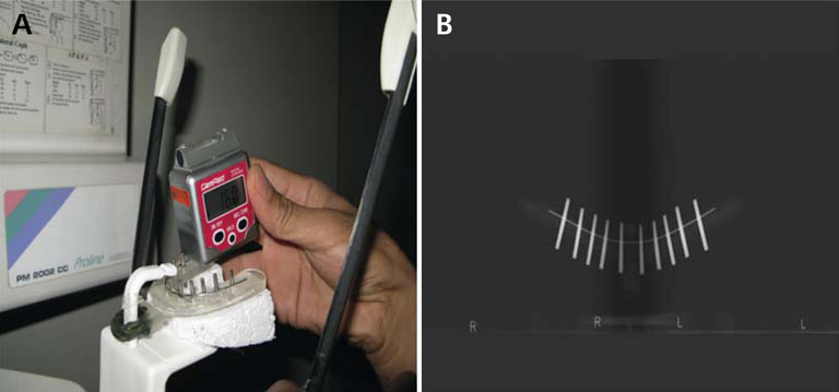
Fig. 3 A. Acrylic model along with the inserted pins in the panoramic machine; B. Panoramic image of the inserted pins in the acrylic model.
Cited by 1 articles
-
Evaluation of factors influencing the success rate of orthodontic microimplants using panoramic radiographs
Jae Hyun Park, Jong-Moon Chae, R. Curtis Bay, Mi-Jung Kim, Keun-Young Lee, Na-Young Chang
Korean J Orthod. 2018;48(1):30-38. doi: 10.4041/kjod.2018.48.1.30.
Reference
-
1. Siu AS, Chu FC, Li TK, Chow TW, Deng FL. Imaging modalities for preoperative assessment in dental implant therapy: an overview. Hong Kong Dent J. 2010; 7:23–30.2. Brooks SL. Guidelines for radiologic examinations: do we have all the answers yet? Oral Surg Oral Med Oral Pathol Oral Radiol Endod. 1997; 83:523–524.
Article3. Choi BR, Choi DH, Huh KH, Yi WJ, Heo MS, Choi SC, et al. Clinical image quality evaluation for panoramic radiography in Korean dental clinics. Imaging Sci Dent. 2012; 42:183–190.
Article4. Rejebian GP. A statistical correlation of individual tooth size distortions on the orthopantomographic radiograph. Am J Orthod. 1979; 75:525–534.
Article5. Batenburg RH, Stellingsma K, Raghoebar GM, Vissink A. Bone height measurements on panoramic radiographs: the effect of shape and position of edentulous mandibles. Oral Surg Oral Med Oral Pathol Oral Radiol Endod. 1997; 84:430–435.6. White SC, Pharoah MJ. Oral radiology: principles and interpretation. 5th ed. St. Louis: Mosby;2009. p. 677–692.7. Frykholm A, Malmgren O, Sämfors KA, Welander U. Angular measurements in orthopantomography. Dentomaxillofac Radiol. 1977; 6:77–81.
Article8. Laster WS, Ludlow JB, Bailey LJ, Hershey HG. Accuracy of measurements of mandibular anatomy and prediction of asymmetry in panoramic radiographic images. Dentomaxillofac Radiol. 2005; 34:343–349.
Article9. Mckee IW, Glover KE, Williamson PC, Lam EW, Heo G, Major PW. The effect of vertical and horizontal head positioning in panoramic radiography on mesiodistal tooth angulations. Angle Orthod. 2001; 71:442–451.10. Catić A, Celebić A, Valentić-Peruzović M, Catović A, Jerolimov V, Muretić I. Evaluation of the precision of dimensional measurements of the mandible on panoramic radiographs. Oral Surg Oral Med Oral Pathol Oral Radiol Endod. 1998; 86:242–248.11. Langland OE, Langlais RP, Preece JW. Principles of dental imaging. Baltimore: Lippincott Williams & Wilkins;2002. p. 201–218. p. 224–271.
Article12. Stramotas S, Geenty JP, Petocz P, Darendeliler MA. Accuracy of linear and angular measurements on panoramic radiographs taken at various positions in vitro. Eur J Orthod. 2002; 24:43–52.
Article13. Tronje G, Eliasson S, Julin P, Welander U. Image distortion in rotational panoramic radiography. II. Vertical distances. Acta Radiol Diagn (Stockh). 1981; 22:449–455.14. Taguchi A, Tanimoto K, Suei Y, Otani K, Wadamoto M, Akagawa Y, et al. Observer agreement in the assessment of mandibular trabecular bone pattern from panoramic radiographs. Dentomaxillofac Radiol. 1997; 26:90–94.
Article15. Lucchesi M, Wood R, Nortje C. Suitability of the panoramic radiograph for assessment of mesiodistal angulation of teeth in the buccal segments of the mandible. Am J Orthod Dentofacial Orthop. 1988; 94:303–310.
Article16. Hoseini Zarch SH, Bagherpour A, Javadian Langaroodi A, Ahmadian Yazdi A, Safaei A. Evaluation of the accuracy of panoramic radiography in linear measurements of the jaws. Iran J Radiol. 2011; 8:97–102.17. Pfeiffer P, Bewersdorf S, Schmage P. The effect of changes in head position on enlargement of structures during panoramic radiography. Int J Oral Maxillofac Implants. 2012; 27:55–63.18. Ladeira DB, Cruz AD, Almeida SM, Bóscolo FN. Evaluation of the panoramic image formation in different anatomic positions. Braz Dent J. 2010; 21:458–462.
Article19. Hardy TC, Suri L, Stark P. Influence of patient head positioning on measured axial tooth inclination in panoramic radiography. J Orthod. 2009; 36:103–110.
Article20. Sanderink GC, Visser WN, Kramers EW. The origin of a case of severe image distortion in rotational panoramic radiography. Dentomaxillofac Radiol. 1991; 20:169–171.
Article
- Full Text Links
-
- Actions
-
Cited
- CITED
-
- Close
- Share
-
- Similar articles
-
- The comparative study of three-dimensional cephalograms to actual models and conventional lateral cephalograma in linear and angular measurements
- A pilot study of an automated personal identification process: Applying machine learning to panoramic radiographs
- Evaluation of mesiodistal tooth axis using a CBCT-generated panoramic view
- Radiographic evaluation of the course and visibility of the mandibular canal
- Changes of lateral cephalometric values according to the rotation of head
