Radiographic evaluation of the course and visibility of the mandibular canal
- Affiliations
-
- 1Department of Oral and Maxillofacial Radiology, School of Dentistry, Pusan National University, Yangsan, Korea. bhjo@pusan.ac.kr
- KMID: 1974495
- DOI: http://doi.org/10.5624/isd.2014.44.4.273
Abstract
- PURPOSE
This study was performed to investigate the course of the mandibular canal on panoramic radiography and the visibility of this canal on both panoramic radiography and cone-beam computed tomography (CBCT).
MATERIALS AND METHODS
The study consisted of panoramic radiographs and CBCT images from 262 patients. The course of the mandibular canal, as seen in panoramic radiographs, was classified into four types: linear, elliptical, spoon-shaped, and turning curves. The visibility of this canal from the first to the third molar region was evaluated by visually determining whether the mandibular canal was clearly visible, probably visible, or invisible. The visibihlity of the canal on panoramic radiographs was compared with that on CBCT images.
RESULTS
Elliptical curves were most frequently observed along the course of the mandibular canal. The percentage of clearly visible mandibular canals was the highest among the spoon-shaped curves and the lowest among the linear curves. On panoramic radiographs, invisible mandibular canals were found in 22.7% of the examined sites in the first molar region, 11.8% in the second molar region, and 1.3% in the third molar region. On CBCT cross-sectional images, the mandibular canal was invisible in 8.2% of the examined sites in the first molar region, 5.7% in the second molar region, and 0.2% in the third molar region.
CONCLUSION
The visibility of this canal was lower in the first molar region than in the third molar region. The mandibular canal presented better visibility on CBCT images than on panoramic radiographs.
MeSH Terms
Figure
-

Fig. 1 Classifications of the course of the mandibular canal on panoramic radiographs: A. Linear curve, the canal curve is approximately a straight line; B. Elliptic curve, the curve is approximately symmetrical; C. Spoon-shaped curve, the canal has an approximate spoon shape that is similar to an asymmetric elliptic arc; and D. Turning curve, the course is unsmooth and has a turning point.
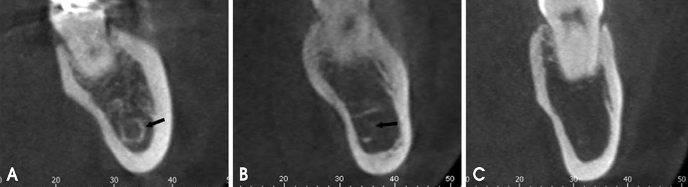
Fig. 2 Classification of the visibility of the mandibular canal on cross-sectional cone-beam computed tomography images: A. Clearly visible, the whole circumference of the bony wall is clearly visible; B. Probably visible, the canal wall is partly visible; and C. Invisible, the canal is not identifiable with certainty.
Cited by 1 articles
-
Common conditions associated with mandibular canal widening: A literature review
Hamed Mortazavi, Maryam Baharvand, Yaser Safi, Kazem Dalaie, Mohammad Behnaz, Fatemeh Safari
Imaging Sci Dent. 2019;49(2):87-95. doi: 10.5624/isd.2019.49.2.87.
Reference
-
1. Oliveira-Santos C, Capelozza AL, Dezzoti MS, Fischer CM, Poleti ML, Rubira-Bullen IR. Visibility of the mandibular canal on CBCT cross-sectional images. J Appl Oral Sci. 2011; 19:240–243.
Article2. Escoda-Francoli J, Canalda-Sahli C, Soler A, Figueiredo R, Gay-Escoda C. Inferior alveolar nerve damage because of overextended endodontic material: a problem of sealer cement biocompatibility? J Endod. 2007; 33:1484–1489.
Article3. Tsuji Y, Muto T, Kawakami J, Takeda S. Computed tomographic analysis of the position and course of the mandibular canal: relevance to the sagittal split ramus osteotomy. Int J Oral Maxillofac Surg. 2005; 34:243–246.
Article4. Kamrun N, Tetsumura A, Nomura Y, Yamaguchi S, Baba O, Nakamura S, et al. Visualization of the superior and inferior borders of the mandibular canal: a comparative study using digital panoramic radiographs and cross-sectional computed tomography images. Oral Surg Oral Med Oral Pathol Oral Radiol. 2013; 115:550–557.
Article5. Worthington P. Injury to the inferior alveolar nerve during implant placement: a formula for protection of the patient and clinician. Int J Oral Maxillofac Implants. 2004; 19:731–734.6. Kieser JA, Paulin M, Law B. Intrabony course of the inferior alveolar nerve in the edentulous mandible. Clin Anat. 2004; 17:107–111.
Article7. Nortjé CJ, Farman AG, Grotepass FW. Variations in the normal anatomy of the inferior dental (mandibular) canal: a retrospective study of panoramic radiographs from 3612 routine dental patients. Br J Oral Surg. 1977; 15:55–63.
Article8. Anderson LC, Kosinski TF, Mentag PJ. A review of the intraosseous course of the nerves of the mandible. J Oral Implantol. 1991; 17:394–403.9. Carter RB, Keen EN. The intramandibular course of the inferior alveolar nerve. J Anat. 1971; 108:433–440.10. Denio D, Torabinejad M, Bakland LK. Anatomical relationship of the mandibular canal to its surrounding structures in mature mandibles. J Endod. 1992; 18:161–165.
Article11. Wadu SG, Penhall B, Townsend GC. Morphological variability of the human inferior alveolar nerve. Clin Anat. 1997; 10:82–87.
Article12. Gowgiel JM. The position and course of the mandibular canal. J Oral Implantol. 1992; 18:383–385.13. Başa O, Dilek OC. Assessment of the risk of perforation of the mandibular canal by implant drill using density and thickness parameters. Gerodontology. 2011; 28:213–220.
Article14. de Oliveira-Santos C, Souza PH, de Azambuja Berti-Couto S, Stinkens L, Moyaert K, Rubira-Bullen IR, et al. Assessment of variations of the mandibular canal through cone beam computed tomography. Clin Oral Investig. 2012; 16:387–393.
Article15. Klinge B, Petersson A, Maly P. Location of the mandibular canal: comparison of macroscopic findings, conventional radiography, and computed tomography. Int J Oral Maxillofac Implants. 1989; 4:327–332.16. Lindh C, Petersson A, Klinge B. Visualisation of the mandibular canal by different radiographic techniques. Clin Oral Implants Res. 1992; 3:90–97.
Article17. Ylikontiola L, Moberg K, Huumonen S, Soikkonen K, Oikarinen K. Comparison of three radiographic methods used to locate the mandibular canal in the buccolingual direction before bilateral sagittal split osteotomy. Oral Surg Oral Med Oral Pathol Oral Radiol Endod. 2002; 93:736–742.
Article18. Kim EK. Comparison of different radiographic methods for the detection of the mandibular canal. Korean J Oral Maxillofac Radiol. 2003; 33:199–205.19. Angelopoulos C, Thomas SL, Hechler S, Parissis N, Hlavacek M. Comparison between digital panoramic radiography and cone-beam computed tomography for the identification of the mandibular canal as part of presurgical dental implant assessment. J Oral Maxillofac Surg. 2008; 66:2130–2135.
Article20. Lofthag-Hansen S, Gröndahl K, Ekestubbe A. Cone-beam CT for preoperative implant planning in the posterior mandible: visibility of anatomic landmarks. Clin Implant Dent Relat Res. 2009; 11:246–255.
Article21. Monsour PA, Dudhia R. Implant radiography and radiology. Aust Dent J. 2008; 53:Suppl 1. S11–S25.
Article22. Ozturk A, Potluri A, Vieira AR. Position and course of the mandibular canal in skulls. Oral Surg Oral Med Oral Pathol Oral Radiol. 2012; 113:453–458.
Article23. Liu T, Xia B, Gu Z. Inferior alveolar canal course: a radiographic study. Clin Oral Implants Res. 2009; 20:1212–1218.
Article24. Heasman PA. Variation in the position of the inferior dental canal and its significance to restorative dentistry. J Dent. 1988; 16:36–39.
Article25. Naitoh M, Katsumata A, Kubota Y, Hayashi M, Ariji E. Relationship between cancellous bone density and mandibular canal depiction. Implant Dent. 2009; 18:112–118.
Article26. Lindh C, Petersson A, Klinge B. Measurements of distances related to the mandibular canal in radiographs. Clin Oral Implants Res. 1995; 6:96–103.
Article27. Bertl K, Heimel P, Reich KM, Schwarze UY, Ulm C. A histomorphometric analysis of the nature of the mandibular canal in the anterior molar region. Clin Oral Investig. 2014; 18:41–47.
Article
- Full Text Links
-
- Actions
-
Cited
- CITED
-
- Close
- Share
-
- Similar articles
-
- Comparison of different radiographic methods for the detection of the mandibular canal
- The effect of different radiographic parameters on the height, width and visibility of cross-sectional image of mandible in spiral tomography
- Analysis and evaluation of relative positions of mandibular third molar and mandibular canal impacts
- Visibility of the mandibular canal and the mental foramen in panoramic radiography
- Reliability of panoramic radiography in predicting proximity of third molars to the mandibular canal: A comparison using cone-beam computed tomography
