Hepatic Metastases of Gastric Adenocarcinoma Showing Metabolic Remission on FDG-PET Despite an Increase in Size on CT
- Affiliations
-
- 1Department of Hematooncology, Konkuk University Medical Center, Seoul, Korea. greenteamd@gmail.com
- 2Department of Nuclear Medicine, Konkuk University Medical Center, Seoul, Korea.
Abstract
- We report a gastric adenocarcinoma patient with liver metastases. The metastases showed progression on computed tomography (CT), but this was not true progression in terms of metabolic activity according to (18)F-fluorodeoxyglucose positron emission tomography (FDG-PET). Discordance between size criteria and metabolic criteria has been reported in liver gastrointestinal stromal tumors, hepatomas, and renal cell carcinomas after dramatic responses with targeted therapies such as imatinib, sorafenib, and sunitinib (1-6). However, this discordance has been rarely reported in liver metastases of gastric adenocarcinoma when treated with conventional chemotherapy.
MeSH Terms
-
Adenocarcinoma
Benzamides
Carcinoma, Hepatocellular
Carcinoma, Renal Cell
Gastrointestinal Stromal Tumors
Humans
Indoles
Liver
Neoplasm Metastasis
Niacinamide
Phenylurea Compounds
Piperazines
Positron-Emission Tomography
Pyrimidines
Pyrroles
Stomach Neoplasms
Imatinib Mesylate
Benzamides
Indoles
Niacinamide
Phenylurea Compounds
Piperazines
Pyrimidines
Pyrroles
Figure
-
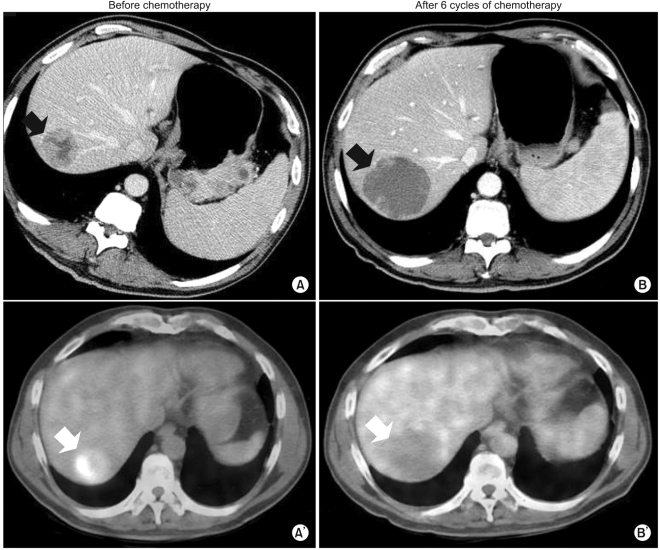
Fig. 1 (A, A') Baseline CT and FDG-PET before chemotherapy. (B, B') CT and FDG-PET after 6 cycles of chemotherapy. After 6 cycles of chemotherapy, the sizes of the metastatic liver masses definitively increased 20% above the original tumor sizes according to CT. This patient showed disease progression according to RECIST criteria. However, the hepatic masses also turned cystic and necrotic compared to the original masses. In addition to that finding on CT, there was no metabolic activity in the liver masses on FDG-PET after chemotherapy.
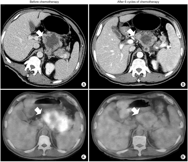
Fig. 2 (A, A') Baseline CT and FDG-PET before chemotherapy. (B, B') CT and FDG-PET after 6 cycles of chemotherapy. The maximum SUV of the lymph nodes decreased from 7.6 into 2.4. Taken together, the patient showed metabolic PR according to maximum SUV on FDG-PET.
Reference
-
1. Abou-Alfa GK, Schwartz L, Ricci S, Amadori D, Santoro A, Figer A, et al. Phase II study of sorafenib in patients with advanced hepatocellular carcinoma. J Clin Oncol. 2006; 24:4293–4300. PMID: 16908937.
Article2. Benjamin RS, Choi H, Macapinlac HA, Burgess MA, Patel SR, Chen LL, et al. We should desist using RECIST, at least in GIST. J Clin Oncol. 2007; 25:1760–1764. PMID: 17470866.
Article3. Choi H, Charnsangavej C, Faria SC, Macapinlac HA, Burgess MA, Patel SR, et al. Correlation of computed tomography and positron emission tomography in patients with metastatic gastrointestinal stromal tumor treated at a single institution with imatinib mesylate: proposal of new computed tomography response criteria. J Clin Oncol. 2007; 25:1753–1759. PMID: 17470865.
Article4. Motzer RJ, Bukowski RM. Targeted therapy for metastatic renal cell carcinoma. J Clin Oncol. 2006; 24:5601–5608. PMID: 17158546.
Article5. Antoch G, Kanja J, Bauer S, Kuehl H, Renzing-Koehler K, Schuette J, et al. Comparison of PET, CT, and dual-modality PET/CT imaging for monitoring of imatinib (STI571) therapy in patients with gastrointestinal stromal tumors. J Nucl Med. 2004; 45:357–365. PMID: 15001674.6. Goerres GW, Stupp R, Barghouth G, Hany TF, Pestalozzi B, Dizendorf E, et al. The value of PET, CT and in-line PET/CT in patients with gastrointestinal stromal tumours: long-term outcome of treatment with imatinib mesylate. Eur J Nucl Med Mol Imaging. 2005; 32:153–162. PMID: 15690223.
Article7. Therasse P, Arbuck SG, Eisenhauer EA, Wanders J, Kaplan RS, Rubinstein L, et al. New guidelines to evaluate the response to treatment in solid tumors. European organization for research and treatment of cancer, national cancer institute of the United States, national cancer institute of Canada. J Natl Cancer Inst. 2000; 92:205–216. PMID: 10655437.
- Full Text Links
-
- Actions
-
Cited
- CITED
-
- Close
- Share
-
- Similar articles
-
- Lung Adenocarcinoma Staged as an Unknown Primary Presenting with Symptomatic Colon Metastases: Staging by 18F-FDG PET/CT
- Imaging of Gastric Cancer Metabolism Using 18 F-FDG PET/CT
- Value of Bone Scan in Addition to F-18 FDG PET/CT and Characteristics of Discordant Lesions between F-18 FDG PET/CT and Bone Scan in the Spinal Bony Metastasis
- Discrepancy of Bone Metastases between F-18 FDG PET/CT and Bone Scan in a Patient with Prostate Cancer
- ¹â¸F-FDG PET/MR Refines Evaluation in Newly Diagnosed Metastatic Urethral Adenocarcinoma
