Recent Advances in Molecular Imaging of Premalignant Gastrointestinal Lesions and Future Application for Early Detection of Barrett Esophagus
- Affiliations
-
- 1Digestive Disease Center, CHA Bundang Medical Center, CHA University, Seongnam, Korea. hahmkb@cha.ac.kr
- 2Gachon University College of Pharmacy, Incheon, Korea.
- 3Cancer Prevention Research Center, CHA University, Seoul, Korea.
Abstract
- Recent advances in optical molecular imaging allow identification of morphologic and biochemical changes in tissues associated with gastrointestinal (GI) premalignant lesions earlier and in real-time. This focused review series introduces high-resolution imaging modalities that are being evaluated preclinically and clinically for the detection of early GI cancers, especially Barrett esophagus and esophageal adenocarcinoma. Although narrow band imaging, autofluorescence imaging, and chromoendoscopy are currently applied for this purpose in the clinic, further adoptions of probe-based confocal laser endomicroscopy, high-resolution microendoscopy, optical coherence tomography, and metabolomic imaging, as well as imaging mass spectrometry, will lead to detection at the earliest and will guide predictions of the clinical course in the near future in a manner that is beyond current advancements in optical imaging. In this review article, the readers will be introduced to sufficient information regarding this matter with which to enjoy this new era of high technology and to confront science in the field of molecular medical imaging.
Keyword
MeSH Terms
Figure
-
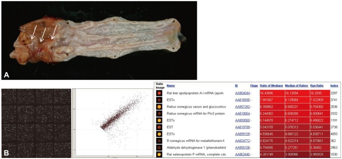
Fig. 1 (A) Animal model for Barrett esophagus (BE). Esophagojejunostomy was performed in Sprague Dawley rats to expose the esophagus to gastroduodenal contents. Histological examination showed the clear appearance of BE with partial changes of BE-associated adenocarcinoma (arrows). (B) cDNA microarray for biomarkers of BE. A 20,000 rat cDNA microarray (Macrogen) was probed using Cy3 and Cy5 labeling to identify the genes responsible for BE and BE-associated carcinoma with Scatchard plotting and bioinformatics analysis. The results are currently being validated.
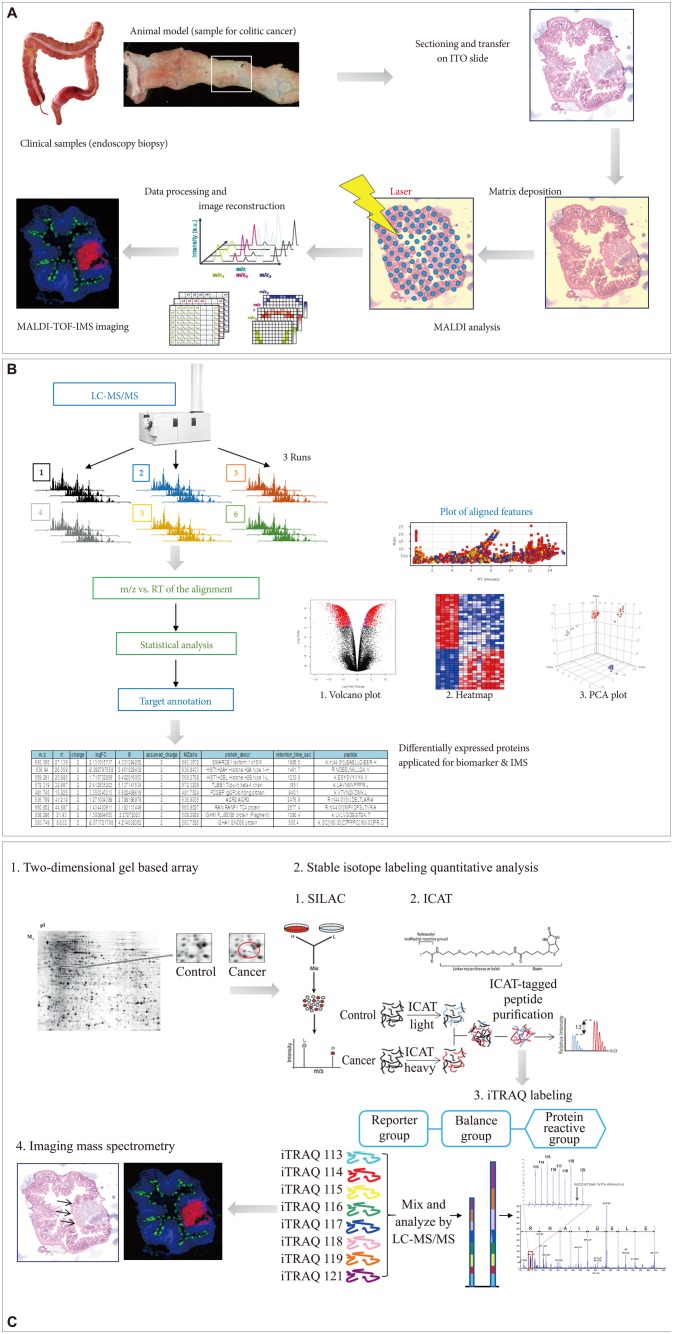
Fig. 2 Advances in molecular imaging technology for future medicine in gastroenterology, and cDNA microarray and imaging mass spectrometry (IMS). (A) Flow for IMS as exemplified in colitic cancer. (B) Label-free protein quantification scheme for either biomarker discovery or IMS. (C) Label-based protein quantification scheme using isobaric tags for relative and absolute quantification (iTRAQ) labeling. ITO, indium tin oxide; MALDI-TOF-IMS, matrix-assisted laser desorption/ionization time-of-flight imaging mass spectrometry; LC-MS/MS, liquid chromatography-tandem mass spectrometry; RT, chromatographic retention time; m/z, mass-to-charge ratio; PCA, principal components analysis.
Reference
-
1. Subramanian V, Ragunath K. Advanced endoscopic imaging: a review of commercially available technologies. Clin Gastroenterol Hepatol. 2013; 6. 28. Epub. DOI:10.1016/j.cgh.2013.06.015.
Article2. Chandra S, Gorospe EC, Leggett CL, Wang KK. Barrett's esophagus in 2012: updates in pathogenesis, treatment, and surveillance. Curr Gastroenterol Rep. 2013; 15:322. PMID: 23605564.
Article3. di Pietro M, Fitzgerald RC. Screening and risk stratification for Barrett's esophagus: how to limit the clinical impact of the increasing incidence of esophageal adenocarcinoma. Gastroenterol Clin North Am. 2013; 42:155–173. PMID: 23452636.4. Qumseya BJ, Wang H, Badie N, et al. Advanced imaging technologies increase detection of dysplasia and neoplasia in patients with Barrett's esophagus: a meta-analysis and systematic review. Clin Gastroenterol Hepatol. 2013; 11:1562–1570. PMID: 23851020.
Article5. Sturm MB, Joshi BP, Lu S, et al. Targeted imaging of esophageal neoplasia with a fluorescently labeled peptide: first-in-human results. Sci Transl Med. 2013; 5:184ra61.
Article6. von Holzen U, Enders GH. A surprise cell of origin for Barrett's esophagus. Cancer Biol Ther. 2012; 13:588–591. PMID: 22549156.
Article7. Franks I. Barrett esophagus: new insights into the stem cell organization of Barrett esophagus. Nat Rev Gastroenterol Hepatol. 2012; 9:125. PMID: 22290494.8. Templeton A, Hwang JH. Confocal microscopy in the esophagus and stomach. Clin Endosc. 2013; 46:445–449. PMID: 24143300.
Article9. Gordon LG, Mayne GC. Cost-effectiveness of Barrett's oesophagus screening and surveillance. Best Pract Res Clin Gastroenterol. 2013; 27:893–903. PMID: 24182609.
Article10. Iwaya Y, Hasebe O, Koide N, et al. Reduced expression of alphaGlcNAc in Barrett's oesophagus adjacent to Barrett's adenocarcinoma - a possible biomarker to predict the malignant potential of Barrett's oesophagus. Histopathology. Epub 2013 Oct 1. DOI: 10.1111/his.12296.11. Kastelein F, Biermann K, Steyerberg EW, et al. Value of alpha-methylacyl-CoA racemase immunochemistry for predicting neoplastic progression in Barrett's oesophagus. Histopathology. 2013; 63:630–639. PMID: 24004067.12. Fels Elliott DR, Fitzgerald RC. Molecular markers for Barrett's esophagus and its progression to cancer. Curr Opin Gastroenterol. 2013; 29:437–445. PMID: 23689523.
Article13. Hasina R, Mollberg N, Kawada I, et al. Critical role for the receptor tyrosine kinase EPHB4 in esophageal cancers. Cancer Res. 2013; 73:184–194. PMID: 23100466.
Article14. Li M, Anastassiades CP, Joshi B, et al. Affinity peptide for targeted detection of dysplasia in Barrett's esophagus. Gastroenterology. 2010; 139:1472–1480. PMID: 20637198.
Article15. Kiesslich R, Gossner L, Goetz M, et al. In vivo histology of Barrett's esophagus and associated neoplasia by confocal laser endomicroscopy. Clin Gastroenterol Hepatol. 2006; 4:979–987. PMID: 16843068.
Article16. Liebler DC, Zimmerman LJ. Targeted quantitation of proteins by mass spectrometry. Biochemistry. 2013; 52:3797–3806. PMID: 23517332.
Article17. Boja ES, Rodriguez H. Mass spectrometry-based targeted quantitative proteomics: achieving sensitive and reproducible detection of proteins. Proteomics. 2012; 12:1093–1110. PMID: 22577011.
Article18. Picotti P, Aebersold R. Selected reaction monitoring-based proteomics: workflows, potential, pitfalls and future directions. Nat Methods. 2012; 9:555–566. PMID: 22669653.
Article19. Liu YW, Aciego SM, Wanamaker AD Jr, Sell BK. A high-throughput system for boron microsublimation and isotope analysis by total evaporation thermal ionization mass spectrometry. Rapid Commun Mass Spectrom. 2013; 27:1705–1714. PMID: 23821564.
Article20. van den Broek I, Sparidans RW, Schellens JH, Beijnen JH. Sensitive liquid chromatography/tandem mass spectrometry assay for absolute quantification of ITIH4-derived putative biomarker peptides in clinical serum samples. Rapid Commun Mass Spectrom. 2010; 24:1842–1850. PMID: 20533314.
Article21. Su Y, Zhu Y, Fang Q. A multifunctional microfluidic droplet-array chip for analysis by electrospray ionization mass spectrometry. Lab Chip. 2013; 13:1876–1882. PMID: 23525283.
Article22. Qiu L, Turzhitsky V, Chuttani R, et al. Spectral imaging with scattered light: from early cancer detection to cell biology. IEEE J Sel Top Quantum Electron. 2012; 18:1073–1083. PMID: 23087592.
Article23. Perelman LT, Backman V, Wallace M, et al. Observation of periodic fine structure in reflectance from biological tissue: a new technique for measuring nuclear size distribution. Phys Rev Lett. 1998; 80:627–630.
Article24. Bedard N, Hagen N, Gao L, Tkaczyk TS. Image mapping spectrometry: calibration and characterization. Opt Eng. 2012; 51:pii: 111711.
Article25. Altelaar AF, Munoz J, Heck AJ. Next-generation proteomics: towards an integrative view of proteome dynamics. Nat Rev Genet. 2013; 14:35–48. PMID: 23207911.
Article26. Stunnenberg HG, Hubner NC. Genomics meets proteomics: identifying the culprits in disease. Hum Genet. 2013; 10. 18. Epub. DOI: 10.1007/s00439-013-1376-2.
Article27. Cramer R. Editorial for "advances in biological mass spectrometry and proteomics". Methods. 2011; 54:349–350. PMID: 21839395.
Article28. Filiou MD, Martins-de-Souza D, Guest PC, Bahn S, Turck CW. To label or not to label: applications of quantitative proteomics in neuroscience research. Proteomics. 2012; 12:736–747. PMID: 22247077.
Article29. Sjödin MO, Wetterhall M, Kultima K, Artemenko K. Comparative study of label and label-free techniques using shotgun proteomics for relative protein quantification. J Chromatogr B Analyt Technol Biomed Life Sci. 2013; 928:83–92.
Article30. Han NY, Choi W, Park JM, Kim EH, Lee H, Hahm KB. Label-free quantification for discovering novel biomarkers in the diagnosis and assessment of disease activity in inflammatory bowel disease. J Dig Dis. 2013; 14:166–174. PMID: 23320753.
Article31. Matsumura CY, Menezes de Oliveira B, Durbeej M, Marques MJ. Isobaric tagging-based quantification for proteomic analysis: a comparative study of spared and affected muscles from mice at the early phase of dystrophy. PLoS One. 2013; 8:e65831. PMID: 23823696.
- Full Text Links
-
- Actions
-
Cited
- CITED
-
- Close
- Share
-
- Similar articles
-
- Confocal Laser Endomicroscopy and Molecular Imaging in Barrett Esophagus and Stomach
- Current Status of Mucosal Imaging with Narrow-Band Imaging in the Esophagus
- Treatment of Barrett's Esophagus
- Role of linked color imaging for upper gastrointestinal disease: present and future
- Usefulness and Future Prospects of Confocal Laser Endomicroscopy for Gastric Premalignant and Malignant Lesions
