Comparative analysis of carrier systems for delivering bone morphogenetic proteins
- Affiliations
-
- 1Department of Dental Hygiene, Eulji University College of Health Science, Seongnam, Korea.
- 2Department of Periodontology, Research Institute for Periodontal Regeneration, Yonsei University College of Dentistry, Seoul, Korea. shchoi726@yuhs.ac
- 3Department of Periodontology, Kyung Hee University School of Dentistry, Seoul, Korea.
- KMID: 2164342
- DOI: http://doi.org/10.5051/jpis.2015.45.4.136
Abstract
- PURPOSE
The objective of this study was to comparatively assess the bone regenerative capacity of absorbable collagen sponge (ACS), biphasic calcium phosphate block (BCP) and collagenated biphasic calcium phosphate (CBCP) loaded with a low dose of recombinant human bone morphogenetic protein-2 (rhBMP-2).
METHODS
The CBCP was characterized by X-ray diffraction and scanning electron microscopy. In rabbit calvaria, four circular 8-mm-diameter defects were created and assigned to one of four groups: (1) blood-filled group (control), (2) rhBMP-2-soaked absorbable collagen sponge (0.05 mg/mL, 0.1 mL; CS group), (3) rhBMP-2-loaded BCP (BCP group), or (4) rhBMP-2-loaded CBCP (CBCP group). The animals were sacrificed either 2 weeks or 8 weeks postoperatively. Histological and histomorphometric analyses were performed.
RESULTS
The CBCP showed web-like collagen fibrils on and between particles. Greater dimensional stability was observed in the BCP and CBCP groups than in the control and the CS groups at 2 and 8 weeks. The new bone formation was significantly greater in the BCP and CBCP groups than in the control and CS groups at 2 weeks, but did not significantly differ among the four groups at 8 week. The CBCP group exhibited more new bone formation in the intergranular space and in the center of the defect compared to the BCP group at 2 weeks, but a similar histologic appearance was observed in both groups at 8 weeks.
CONCLUSIONS
The dose of rhBMP-2 in the present study enhanced bone regeneration in the early healing period when loaded on BCP and CBCP in rabbit calvarial defects.
MeSH Terms
Figure
-

Figure 1 Clinical photograph of the surgical procedure. The 8-mm-diameter defects were filled with blood coagulum (control), recombinant human bone morphogenetic protein (rhBMP-2)-loaded absorbable collagen sponge (CS), rhBMP-2-loaded biphasic calcium phosphate (BCP), or rhBMP-2-loaded collagenated BCP (CBCP).

Figure 2 X-ray diffraction patterns of collagenated biphasic calcium phosphate (CBCP). The X-ray diffraction spectrum of the CBCP corresponded with the composite phases of hydroxyapatite and β-tricalcium phosphate.

Figure 3 Gross view and microstructure of biphasic calcium phosphate (BCP) and collagenated BCP (CBCP). (A) Clinical photographs in wet or dry conditions of BCP and CBCP. (B) Scanning electron microscopy views of BCP and CBCP.
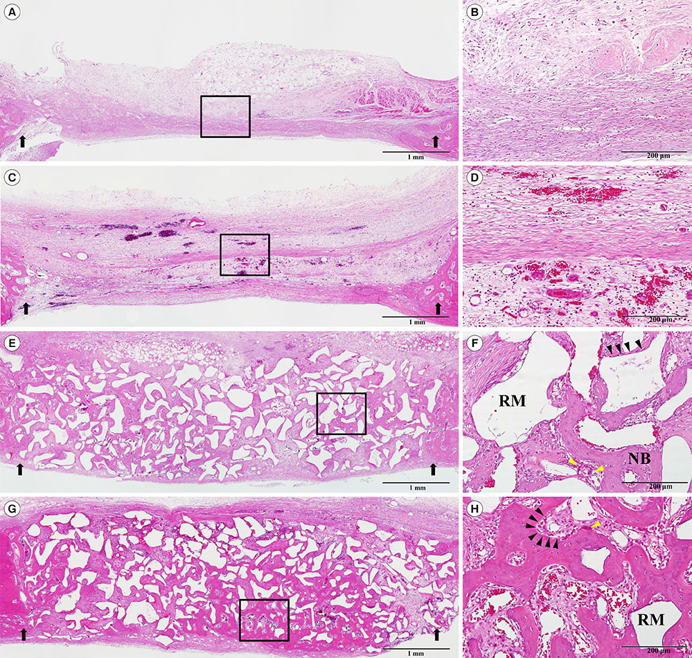
Figure 4 Histologic observations at 2 weeks postsurgery. (A, B) the control group, (C, D) the collagen sponge (CS) group, (E, F) the biphasic calcium phosphate (BCP) group, (G, H) the collagenated BCP (CBCP) group. (B, D, G, F) Higher magnification views of the boxed area in A, C, E, and G, respectively. The black arrows indicate the margins of the defect. The black and yellow arrowheads indicate osteoblasts and osteoclasts, respectively. Hematoxylin-eosin stain; scale bar (A, C, E, G)= 1 mm, (B, D, G, F)= 200 µm. NB, newly formed bone; RM, residual material.
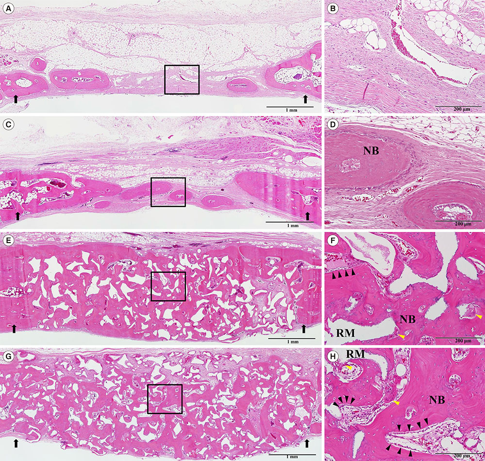
Figure 5 Histologic observations at 8 weeks postsurgery. (A, B) the control group, (C, D) the collagen sponge (CS) group, (E, F) the biphasic calcium phosphate (BCP) group, (G, H) the collagenated BCP (CBCP) group. (B, D, G, F) Higher magnification views of the boxed area in A, C, E, and G, respectively. The black arrows indicate the margins of the defect. The black and yellow arrowheads indicate osteoblasts and osteoclasts, respectively. Hematoxylin-eosin stain; scale bar (A, C, E, G)= 1 mm, (B, D, G, F)= 200 µm. NB, newly formed bone; RM, residual material.

Figure 6 Histomorphometric graphs of the measured parameters. The total length of each bar indicates the total augmented area of each group at each healing period. NB, the area of new bone; FA, the area of fibrovascular tissue; RM, the area of residual material.
Cited by 1 articles
-
Sinus augmentation using rhBMP-2-loaded synthetic bone substitute with simultaneous implant placement in rabbits
Myung-Jae Joo, Jae-Kook Cha, Hyun-Chang Lim, Seong-Ho Choi, Ui-Won Jung
J Periodontal Implant Sci. 2017;47(2):86-95. doi: 10.5051/jpis.2017.47.2.86.
Reference
-
1. Alam I, Asahina I, Ohmamiuda K, Enomoto S. Comparative study of biphasic calcium phosphate ceramics impregnated with rhBMP-2 as bone substitutes. J Biomed Mater Res. 2001; 54:129–138.
Article2. Chang YY, Lee JS, Kim MS, Choi SH, Chai JK, Jung UW. Comparison of collagen membrane and bone substitute as a carrier for rhBMP-2 in lateral onlay graft. Clin Oral Implants Res. 2015; 26:e13–e19.
Article3. Jung RE, Weber FE, Thoma DS, Ehrbar M, Cochran DL, Hämmerle CH. Bone morphogenetic protein-2 enhances bone formation when delivered by a synthetic matrix containing hydroxyapatite/tricalciumphosphate. Clin Oral Implants Res. 2008; 19:188–195.
Article4. Kim JS, Cha JK, Cho AR, Kim MS, Lee JS, Hong JY, et al. Acceleration of Bone Regeneration by BMP-2-Loaded Collagenated Biphasic Calcium Phosphate in Rabbit Sinus. Clin Implant Dent Relat Res. 2014; Forthcoming.
Article5. Kim JW, Choi KH, Yun JH, Jung UW, Kim CS, Choi SH, et al. Bone formation of block and particulated biphasic calcium phosphate lyophilized with Escherichia coli-derived recombinant human bone morphogenetic protein 2 in rat calvarial defects. Oral Surg Oral Med Oral Pathol Oral Radiol Endod. 2011; 112:298–306.
Article6. Lopes NM, Vajgel A, de Oliveira DM, de Santana Santos T, Wassall T. Use of rhBMP-2 to reconstruct a severely atrophic mandible: a modified approach. Int J Oral Maxillofac Surg. 2012; 41:1566–1570.
Article7. Mehanna R, Koo S, Kim DM. Recombinant human bone morphogenetic protein 2 in lateral ridge augmentation. Int J Periodontics Restorative Dent. 2013; 33:97–102.
Article8. Kim YJ, Lee JY, Kim JE, Park JC, Shin SW, Cho KS. Ridge preservation using demineralized bone matrix gel with recombinant human bone morphogenetic protein-2 after tooth extraction: a randomized controlled clinical trial. J Oral Maxillofac Surg. 2014; 72:1281–1290.
Article9. Triplett RG, Nevins M, Marx RE, Spagnoli DB, Oates TW, Moy PK, et al. Pivotal, randomized, parallel evaluation of recombinant human bone morphogenetic protein-2/absorbable collagen sponge and autogenous bone graft for maxillary sinus floor augmentation. J Oral Maxillofac Surg. 2009; 67:1947–1960.
Article10. Carragee EJ, Hurwitz EL, Weiner BK. A critical review of recombinant human bone morphogenetic protein-2 trials in spinal surgery: emerging safety concerns and lessons learned. Spine J. 2011; 11:471–491.
Article11. Kim MS, Lee JS, Shin HK, Kim JS, Yun JH, Cho KS. Prospective randomized, controlled trial of sinus grafting using Escherichia-coli-produced rhBMP-2 with a biphasic calcium phosphate carrier compared to deproteinized bovine bone. Clin Oral Implants Res. 2014; Forthcoming.
Article12. Choi Y, Lee JS, Kim YJ, Kim MS, Choi SH, Cho KS, et al. Recombinant human bone morphogenetic protein-2 stimulates the osteogenic potential of the Schneiderian membrane: a histometric analysis in rabbits. Tissue Eng Part A. 2013; 19:1994–2004.
Article13. Leknes KN, Yang J, Qahash M, Polimeni G, Susin C, Wikesjö UM. Alveolar ridge augmentation using implants coated with recombinant human bone morphogenetic protein-2: radiographic observations. Clin Oral Implants Res. 2008; 19:1027–1033.
Article14. Boerckel JD, Kolambkar YM, Dupont KM, Uhrig BA, Phelps EA, Stevens HY, et al. Effects of protein dose and delivery system on BMP-mediated bone regeneration. Biomaterials. 2011; 32:5241–5251.
Article15. Choi H, Park NJ, Jamiyandorj O, Hong MH, Oh S, Park YB, et al. Improvement of osteogenic potential of biphasic calcium phosphate bone substitute coated with synthetic cell binding peptide sequences. J Periodontal Implant Sci. 2012; 42:166–172.
Article16. Kim JW, Jung IH, Lee KI, Jung UW, Kim CS, Choi SH, et al. Volumetric bone regenerative efficacy of biphasic calcium phosphate-collagen composite block loaded with rhBMP-2 in vertical bone augmentation model of a rabbit calvarium. J Biomed Mater Res A. 2012; 100:3304–3313.
Article17. Kim MS, Kwon JY, Lee JS, Song JS, Choi SH, Jung UW. Low-dose recombinant human bone morphogenetic protein-2 to enhance the osteogenic potential of the Schneiderian membrane in the early healing phase: in vitro and in vivo studies. J Oral Maxillofac Surg. 2014; 72:1480–1494.
Article18. Choi Y, Yun JH, Kim CS, Choi SH, Chai JK, Jung UW. Sinus augmentation using absorbable collagen sponge loaded with Escherichia coli-expressed recombinant human bone morphogenetic protein 2 in a standardized rabbit sinus model: a radiographic and histologic analysis. Clin Oral Implants Res. 2012; 23:682–689.
Article19. Lu SX, Fiorini T, Lee J, Prasad HS, Buxton AN, Bisch FC, et al. Evaluation of a compression resistant matrix for recombinant human bone morphogenetic protein-2. J Clin Periodontol. 2013; 40:688–697.
Article20. McClellan JW, Mulconrey DS, Forbes RJ, Fullmer N. Vertebral bone resorption after transforaminal lumbar interbody fusion with bone morphogenetic protein (rhBMP-2). J Spinal Disord Tech. 2006; 19:483–486.
Article21. Yun PY, Kim YK, Jeong KI, Park JC, Choi YJ. Influence of bone morphogenetic protein and proportion of hydroxyapatite on new bone formation in biphasic calcium phosphate graft: two pilot studies in animal bony defect model. J Craniomaxillofac Surg. 2014; 42:1909–1917.
Article22. Kim DM, Nevins ML, Lin Z, Fateh A, Kim SW, Schupbach P, et al. The clinical and histologic outcome of dental implant in large ridge defect regenerated with alloplast: a randomized controlled preclinical trial. J Oral Implantol. 2013; 39:148–153.
Article23. Nevins M, Nevins ML, Schupbach P, Kim SW, Lin Z, Kim DM. A prospective, randomized controlled preclinical trial to evaluate different formulations of biphasic calcium phosphate in combination with a hydroxyapatite collagen membrane to reconstruct deficient alveolar ridges. J Oral Implantol. 2013; 39:133–139.
Article24. Jang JW, Yun JH, Lee KI, Jang JW, Jung UW, Kim CS, et al. Osteoinductive activity of biphasic calcium phosphate with different rhBMP-2 doses in rats. Oral Surg Oral Med Oral Pathol Oral Radiol. 2012; 113:480–487.
Article25. Yang CR, Wang YJ, Chen XF, Zhao NR. Biomimetic fabrication of BCP/COL/HCA scaffolds for bone tissue engineering. Mater Lett. 2005; 59:3635–3640.
Article26. Brodie JC, Goldie E, Connel G, Merry J, Grant MH. Osteoblast interactions with calcium phosphate ceramics modified by coating with type I collagen. J Biomed Mater Res A. 2005; 73:409–421.
Article27. Brodie JC, Merry J, Grant MH. The mechanical properties of calcium phospate ceramics modified by collagen coating and populated by osteoblasts. J Mater Sci Mater Med. 2006; 17:43–48.
Article28. Hsu EL, Ghodasra JH, Ashtekar A, Nickoli MS, Lee SS, Stupp SI, et al. A comparative evaluation of factors influencing osteoinductivity among scaffolds designed for bone regeneration. Tissue Eng Part A. 2013; 19:1764–1772.
Article29. Seeherman H, Wozney J, Li R. Bone morphogenetic protein delivery systems. Spine (Phila Pa 1976). 2002; 27:S16–S23.
Article30. LeGeros RZ, Lin S, Rohanizadeh R, Mijares D, LeGeros JP. Biphasic calcium phosphate bioceramics: preparation, properties and applications. J Mater Sci Mater Med. 2003; 14:201–209.31. Yang C, Unursaikhan O, Lee JS, Jung UW, Kim CS, Choi SH. Osteoconductivity and biodegradation of synthetic bone substitutes with different tricalcium phosphate contents in rabbits. J Biomed Mater Res B Appl Biomater. 2014; 102:80–88.
Article32. Maté-Sánchez de Val JE, Mazón P, Guirado JL, Ruiz RA, Ramírez Fernández MP, Negri B, et al. Comparison of three hydroxyapatite/beta-tricalcium phosphate/collagen ceramic scaffolds: an in vivo study. J Biomed Mater Res A. 2014; 102:1037–1046.
Article33. Hwang JW, Park JS, Lee JS, Jung UW, Kim CS, Cho KS, et al. Comparative evaluation of three calcium phosphate synthetic block bone graft materials for bone regeneration in rabbit calvaria. J Biomed Mater Res B Appl Biomater. 2012; 100:2044–2052.
Article34. Wikesjö UM, Huang YH, Polimeni G, Qahash M. Bone morphogenetic proteins: a realistic alternative to bone grafting for alveolar reconstruction. Oral Maxillofac Surg Clin North Am. 2007; 19:535–551. vi–vii.
Article35. Montesano R, Orci L, Vassalli P. In vitro rapid organization of endothelial cells into capillary-like networks is promoted by collagen matrices. J Cell Biol. 1983; 97:1648–1652.
Article36. Raines EW. The extracellular matrix can regulate vascular cell migration, proliferation, and survival: relationships to vascular disease. Int J Exp Pathol. 2000; 81:173–182.
Article
- Full Text Links
-
- Actions
-
Cited
- CITED
-
- Close
- Share
-
- Similar articles
-
- The Analysis of Bone regenerative effect with carriers of bone morphogenetic protein in rat calvarial defects
- Purification of porcine bone morphogenetic protein
- The experimental study on the effect of pulsating electromagnetic fields in the osteoinduction induced by bone morphogenetic protein
- Effect of composite of bone morphogenetic protein and plaster of paris on healing of bone defect in the rat tibia
- Effect of bone wax plus bone morphogenetic protein into rabbit calvarial defects
