A Case of Metachronous Development of Esophageal Squamous Cell Carcinoma in the Patient with Esophageal Carcinosarcoma
- Affiliations
-
- 1Department of Internal Medicine, Gyeongsang National University School of Medicine, Jinju, Korea. wtjung@gnu.ac.kr
- 2Department of Pathology, Gyeongsang National University School of Medicine, Jinju, Korea.
- 3Institute of Health Sciences, Gyeongsang National University School of Medicine, Jinju, Korea.
- KMID: 2160669
- DOI: http://doi.org/10.4166/kjg.2014.64.6.364
Abstract
- Esophageal carcinosarcoma is a rare malignant esophageal neoplasm consisting of both carcinomatous and sarcomatous elements, with an incidence of 0.5%. There have been only a few case reports of carcinosarcoma and squamous cell carcinoma coexisting in the esophagus. However, all of these are cases of synchronous or metachronous development of carcinosarcoma after chemoradiotherapy in patients of esophageal squamous cell carcinoma. A 53-year-old man underwent esophagogastroduodenoscopy because of chest pain for several months. Endoscopic examination revealed a huge pedunculated esophageal polypoid mass. Endoscopic submucosal dissection (ESD) was performed and histopathologic examination confirmed spindle cell carcinoma (carcinosarcoma). He refused additional esophagectomy. After 21 months, third follow-up endoscopy showed poorly-demarcated flat, faint discolored lesions at different location from the previous ESD site and endoscopic biopsies confirmed squamous cell carcinoma. To the best of our knowledge, this is the first case of metachronous development of esophageal squamous cell carcinoma in a patient with esophageal carcinosarcoma.
MeSH Terms
-
Antineoplastic Agents/therapeutic use
Carcinoma, Squamous Cell/*diagnosis/drug therapy/pathology
Carcinosarcoma/*diagnosis/drug therapy/pathology
Cisplatin/therapeutic use
Drug Therapy, Combination
Endoscopy, Digestive System
Esophageal Neoplasms/*diagnosis/drug therapy/pathology
Fluorouracil/therapeutic use
Humans
Male
Middle Aged
Positron-Emission Tomography
S100 Proteins/metabolism
Tomography, X-Ray Computed
Tumor Suppressor Protein p53/metabolism
Antineoplastic Agents
Cisplatin
Fluorouracil
S100 Proteins
Tumor Suppressor Protein p53
Figure
-
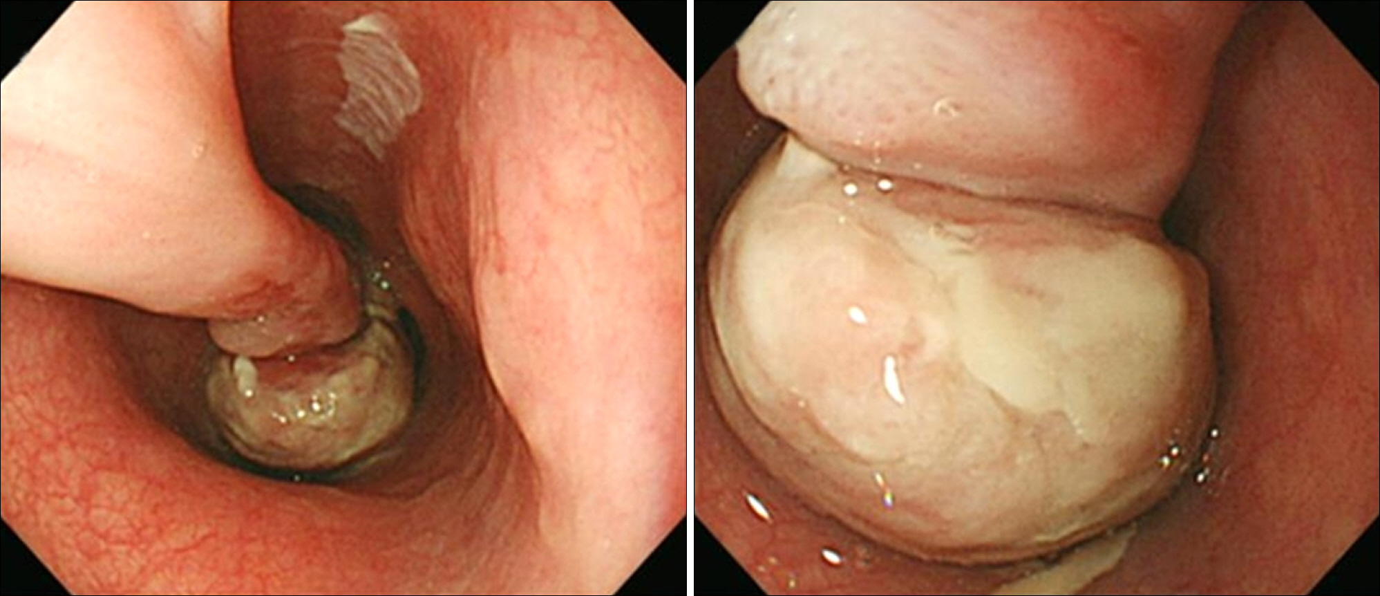
Fig. 1. Endoscopic examination reveals a huge pedunculated esophageal polyp. The polyp stalk originates at 25 cm from the upper incisors and the polyp head extends down to 33 cm.
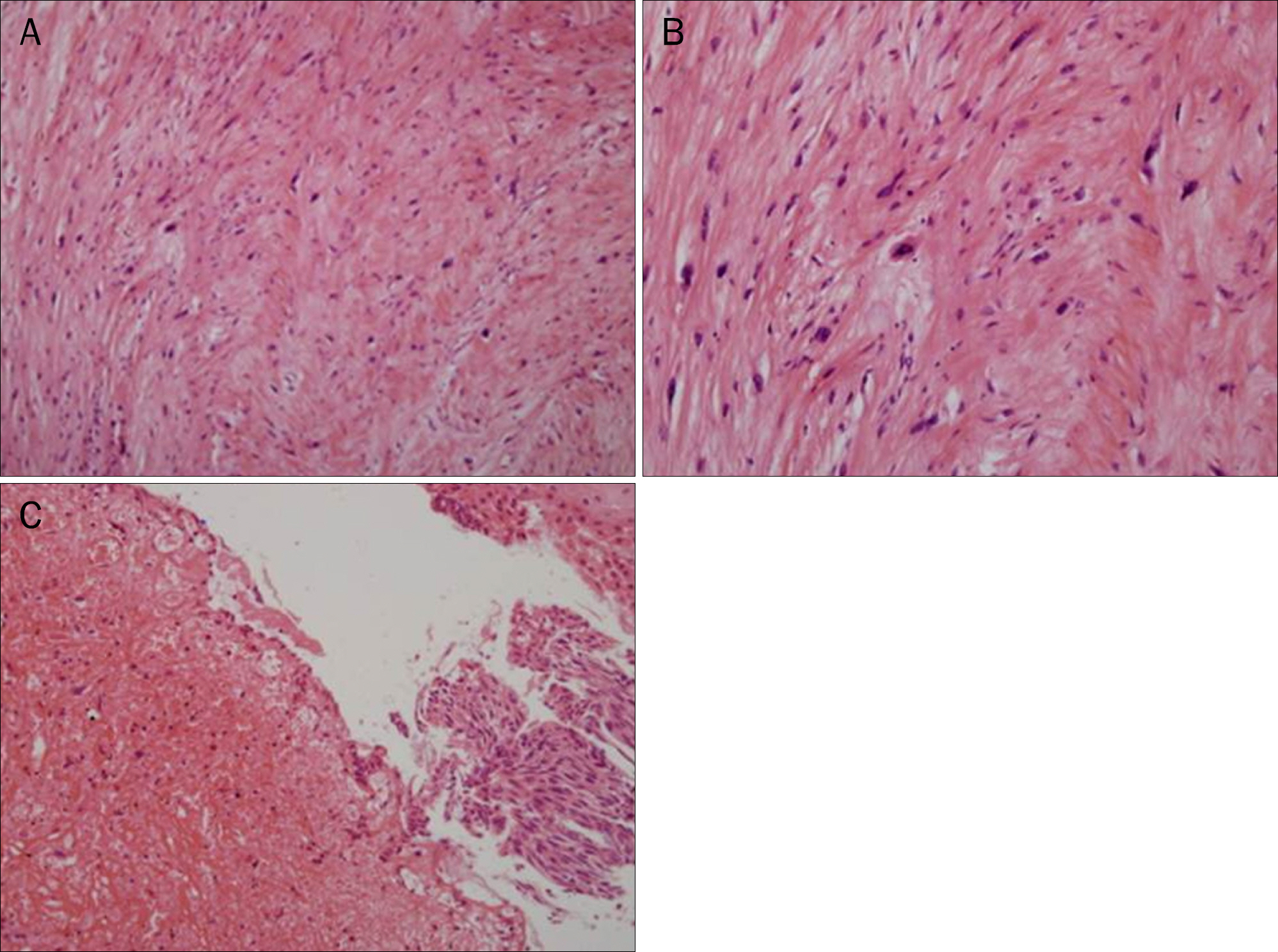
Fig. 2. (A) Histologic finding of a esophageal pedunculated polyp after endoscopic submucosal dissection shows pleomorphic spindle cells and bizarre giant cells with frequent mitotic features (H&E, ×100). (B) The tumor is composed of collagenous connective tissue matrix in which are scattered spindled or pleomorphic tumor cells with hyperchromatic nuclei and frequent mitotic figures (H&E, ×200). (C) There is a very small focus of carcinomatous component (right side of the field), which is a squamous cell carcinoma in situ. Peripheral portion of the sarcomatous component is seen in the left side of the field, which is accompanied by hemorrhage (H&E, ×200).
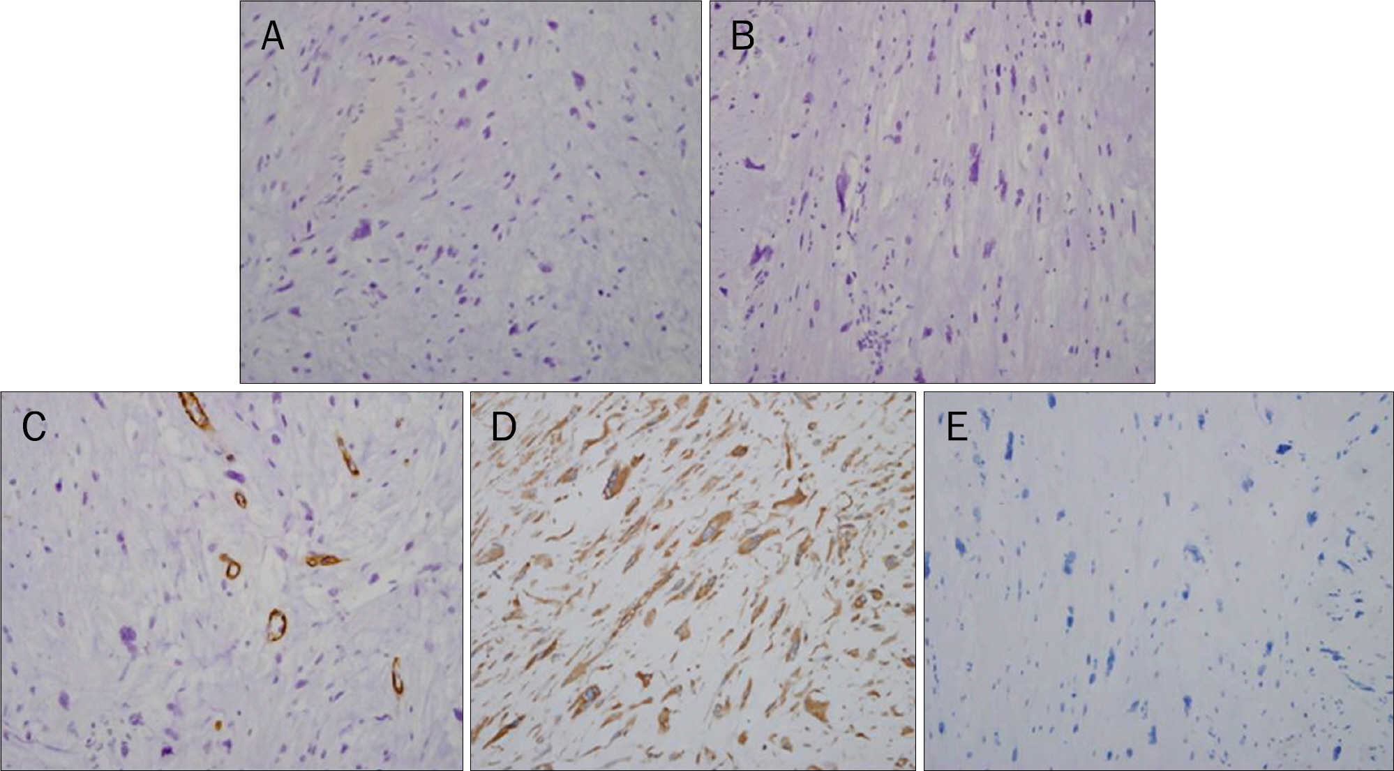
Fig. 3. (A) The tumor cells are negative for cytokeratin (×200). (B) The tumor cells are negative for S-100 protein (×200). (C) The tumor cells are negative for smooth muscle actin. In contrast, normal smooth muscle cells around blood vessels are positive for smooth muscle actin (×200).(D) Many tumor cells are positive for vimetin (×200). (E) The tumor cells are negative for epithelial membrane antigen (×200).
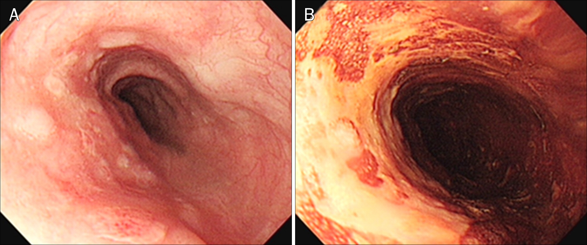
Fig. 4. Twenty-one months later, endoscopic examination demonstrated poorly-demarcated flat, faint discolored lesions at 30–34 cm from the upper incisors (A), which are unstained by Lugol solution (B).

Fig. 5. A forcep biopsy specimen from flat discolored lesions in the esophagus shows well-differentiated squamous cell carcinoma (H&E; A, ×100, B, ×400).
Reference
-
References
1. Iyomasa S, Kato H, Tachimori Y, Watanabe H, Yamaguchi H, Itabashi M. Carcinosarcoma of the esophagus: a twenty-case study. Jpn J Clin Oncol. 1990; 20:99–106.2. Wang ZY, Itabashi M, Hirota T, Watanabe H, Kato H. Immunohistochemical study of the histogenesis of esophageal carcinosarcoma. Jpn J Clin Oncol. 1992; 22:377–386.3. Uchiyama S, Imai S, Hoshino A, et al. Rapid-growing carcinosarcoma of the esophagus arising from intraepithelial squamous cell carcinoma: report of a case. Surg Today. 2000; 30:173–176.
Article4. Zhao S, Xue Q, Ye B, Lu H, He J, Zhao H. Synchronous primary carcinosarcoma and adenosquamous carcinoma of the esophagus. Ann Thorac Surg. 2011; 91:926–928.
Article5. Tomizawa Y, Taniguchi M, Mori M. An unusual case of intra-luminally growing esophageal tumor. Diagnosis: carcinosarcoma of the esophagus. Gastroenterology. 2011; 141:e10–e11.6. Nakagawa S, Yabusaki H, Tanaka O. Rapid-growth carcinosarcoma of the esophagus arising from 0-IIc squamous cell carcinoma after definitive chemoradiotherapy: a case report. Esophagus. 2009; 6:123–126.
Article7. Kuo CJ, Lin TN, Lin CJ, et al. Clinical manifestation of esophageal carcinosarcoma: a Taiwan experience. Dis Esophagus. 2010; 23:122–127.
Article8. Kimura H, Konishi K, Kawamura T, et al. Esophageal sarcomas: report of three cases. Dig Surg. 1999; 16:244–247.
Article9. Madan AK, Long AE, Weldon CB, Jaffe BM. Esophageal carcinosarcoma. J Gastrointest Surg. 2001; 5:414–417.
Article10. Chino O, Kijima H, Shimada H, et al. Clinicopathological studies of esophageal carcinosarcoma: analyses of its morphological characteristics using endoscopic, histological, and immunohistochemical procedures. Endoscopy. 2000; 32:706–711.
Article11. Iascone C, Barreca M. Carcinosarcoma and pseudosarcoma of the esophagus: two names, one disease–comprehensive review of the literature. World J Surg. 1999; 23:153–157.
Article12. Ziauddin MF, Rodriguez HE, Quiros ED, Connolly MM, Podbielski FJ. Carcinosarcoma of the esophagus–pattern of recurrence. Dig Surg. 2001; 18:216–218.13. Ji F, Xu YM, Xu CF. Endoscopic polypectomy: a promising therapeutic choice for esophageal carcinosarcoma. World J Gastroenterol. 2009; 15:3448–3450.
Article14. Sasajima K, Taniguchi Y, Morino K, et al. Rapid growth of a pseudosarcoma of the esophagus. J Clin Gastroenterol. 1988; 10:533–536.15. Pesenti C, Bories E, Danisi C, Monges G, Giovannini M. Endoscopic treatment of esophageal carcinosarcoma: report of a case. Endoscopy. 2004; 36:95.
- Full Text Links
-
- Actions
-
Cited
- CITED
-
- Close
- Share
-
- Similar articles
-
- A Case of Metachronous Carcinosarcoma and Squamous Cell Carcinoma of the Both Parotid Glands
- Cutaneous Metastasis of Esophageal Squamous Cell Carcinoma Mimicking Benign Soft Tissue Tumor
- Carcinosarcoma of the Esophagus: 2 Case Report
- A Case of Early Esophageal Cancer Associated with Invasive Thymoma
- Diagnosis and Clinical Management of Esophageal Squamous Dysplasia
