Ann Dermatol.
2011 May;23(2):205-208. 10.5021/ad.2011.23.2.205.
A Case of Dowling-Degos Disease on the Vulva
- Affiliations
-
- 1Department of Dermatology, Hanyang University Guri Hospital, Hanyang University College of Medicine, Guri, Korea. yuhjoon@hanyang.ac.kr
- KMID: 2156664
- DOI: http://doi.org/10.5021/ad.2011.23.2.205
Abstract
- Dowling-Degos disease (DDD) is an autosomal dominant genodermatosis and this disease is a genetically determined disturbance of epidermal proliferation. It is characterized by acquired, slowly progressive pigmented lesions that primarily involve the great skin folds and flexural areas such as the axilla, neck, limb flexures, the inframammary area and the inguinal folds. The vulva is an unusual location for DDD. A 41-year-old woman presented with a 10-year history of multiple, small, reticulated and brownish macules distributed symmetrically on the bilateral external genital regions. We found no other similarly pigmented skin lesions on her body, including the flexural areas. There was no known family history of similar eruptions or pigmentary changes. The histologic examination showed irregular rete ridge elongation with a filiform or antler-like pattern and basilar hyperpigmentation on the tips. Fontana-Masson staining showed increased pigmentation of the rete ridges and the S100 protein staining did not reveal an increased number of melanocytes in the epidermis. From these findings, we diagnosed this lesion as DDD.
Keyword
MeSH Terms
Figure
-
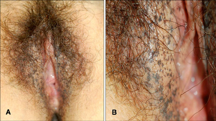
Fig. 1 (A) Multiple hyperpigmented brownish macules located symmetrically on the bilateral external genital area. (B) A close up view of the reticulate pigmented lesion.
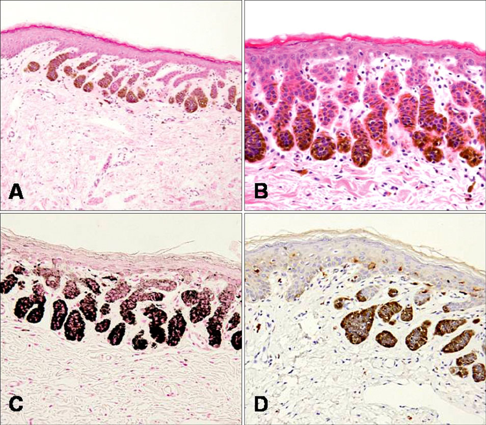
Fig. 2 (A) The epidermal downgrowths showed a filiform or antler-like pattern with irregular elongation of the rete ridges in the lesion (H&E, ×100). (B) There is increased melanin pigment at the tips of the epidermis (H&E, ×200). (C) There is increased pigmentation of the rete ridges on Fontana-Masson staining (Fontana-Masson, ×200). (D) Compared with perilesional normal skin, the S100 protein staining reveals no increase in the number of melanocytes (S100 protein, ×200).
Reference
-
1. Rubio C, Mayor M, Martín MA, González-Beato MJ, Contreras F, Casado M. Atypical presentation of Dowling-Degos disease. J Eur Acad Dermatol Venereol. 2006. 20:1162–1164.
Article2. Massone C, Hofmann-Wellenhof R. Dermoscopy of Dowling-Degos disease of the vulva. Arch Dermatol. 2008. 144:417–418.
Article3. Kim YC, Davis MD, Schanbacher CF, Su WP. Dowling-Degos disease (reticulate pigmented anomaly of the flexures): a clinical and histopathologic study of 6 cases. J Am Acad Dermatol. 1999. 40:462–467.
Article4. Jones EW, Grice K. Reticulate pigmented anomaly of the flexures. Dowing Degos disease, a new genodermatosis. Arch Dermatol. 1978. 114:1150–1157.
Article5. Milde P, Goerz G, Plewig G. Dowling-Degos disease with exclusively genital manifestations. Hautarzt. 1992. 43:369–372.6. O'Goshi K, Terui T, Tagami H. Dowling-Degos disease affecting the vulva. Acta Derm Venereol. 2001. 81:148.7. Wenzel G, Petrow W, Tappe K, Gerdsen R, Uerlich WP, Bieber T. Treatment of Dowling-Degos disease with Er:YAG-laser: results after 2.5 years. Dermatol Surg. 2003. 29:1161–1162.
Article8. Harper JI, Trembath RC. Burns T, Breathnach S, Cox N, Griffiths C, editors. Genetics and genodermatoses. Rook's textbook of dermatology. 2004. 7th ed. Malden: Blackwell;12.82.
Article9. Wu YH, Lin YC. Generalized Dowling-Degos disease. J Am Acad Dermatol. 2007. 57:327–334.
Article10. Betz RC, Planko L, Eigelshoven S, Hanneken S, Pasternack SM, Bussow H, et al. Loss-of-function mutations in the keratin 5 gene lead to Dowling-Degos disease. Am J Hum Genet. 2006. 78:510–519.
Article11. Jeon HJ, Kim SH, Whang KK, Hahm JH. A case of reticulate pigmented anomaly of the flexures (Dowling-Degos disease). Korean J Dermatol. 2006. 44:877–880.12. Howell JB, Freeman RG. Reticular pigmented anomaly of the flexures. Arch Dermatol. 1978. 114:400–403.
Article13. Elder DE, Elenitsas R, Murphy GF, Xu X. Elder DE, Elenitsas R, Johnson BL, Murphy GF, Xu X, editors. Benign pigmented lesions and malignant melanoma. Lever's histopathology of the skin. 2008. 10th ed. Philadelphia: Lippincott-Raven;708–709.14. Juhn BJ, Lee MH, Haw CR. A case of Dowling-Degos disease. Korean J Dermatol. 1999. 37:752–755.15. Alfadley A, Al Ajlan A, Hainau B, Pedersen KT, Al Hoqail I. Reticulate acropigmentation of Dohi: a case report of autosomal recessive inheritance. J Am Acad Dermatol. 2000. 43:113–117.
Article16. Fenske NA, Groover CE, Lober CW, Espinoza CG. Dowling-Degos disease, hidradenitis suppurativa, and multiple keratoacanthomas. A disorder that may be caused by a single underlying defect in pilosebaceous epithelial proliferation. J Am Acad Dermatol. 1991. 24:888–892.
Article17. Wenzel J, Tappe K, Gerdsen R, Uerlich M, Philipp-Dormston W, Bieber T, et al. Successful treatment of Dowling-Degos disease with Er:YAG laser. Dermatol Surg. 2002. 28:748–750.
Article18. Altomare G, Capella GL, Fracchiolla C, Frigerio E. Effectiveness of topical adapalene in Dowling-Degos disease. Dermatology. 1999. 198:176–177.
Article
- Full Text Links
-
- Actions
-
Cited
- CITED
-
- Close
- Share
-
- Similar articles
-
- A Case of Dowling-degos Disease Affecting the Vulva
- A Case of Dowling-Degos Disease
- A Case of Reticulate Pigmented Anomaly of the Flexures (Dowling-Degos Disease)
- A Case of Reticulate Pigmented Anomaly of the Flexures
- A case of reticulate acropigmentation of Kitamura with hyperpigmented macular lesions on the scrotum
