Development of Coronary Vasospasm during Adenosine-Stress Myocardial Perfusion CT Imaging
- Affiliations
-
- 1Department of Radiology, Ulsan University Hospital, University of Ulsan College of Medicine, Ulsan 682-714, Korea. drcsh@naver.com
- KMID: 2155540
- DOI: http://doi.org/10.3348/kjr.2015.16.3.673
Abstract
- Adenosine is a short-acting coronary vasodilator, and it is widely used during pharmacological stress myocardial perfusion imaging. It has a well-established safety profile, and most of its side effects are known to be mild and transient. Until now, coronary vasospasm has been rarely reported as a side effect of adenosine during or after adenosine stress test. This study reports a case of coronary vasospasm which was documented on stress myocardial perfusion CT imaging during adenosine stress test.
MeSH Terms
Figure
-
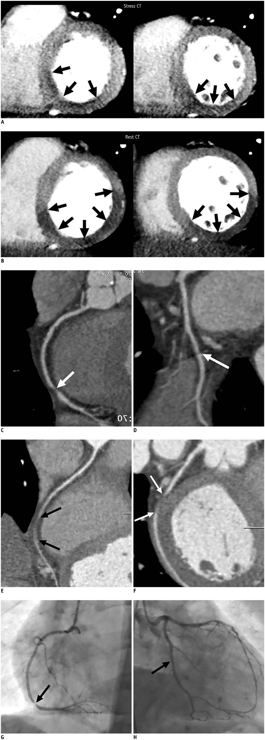
Fig. 1 73-year-old man presenting with chest pain. Stress and rest myocardial perfusion CT images. A. Stress perfusion CT images in short-axis view showed mild to moderate subendocardial perfusion defect in mid to basal inferior and inferoseptal walls of left ventricle (arrows). B. Rest perfusion CT images in short-axis view showed increased extent of myocardial perfusion defect in mid to basal inferior, inferoseptal, and inferolateral walls of left ventricle (arrows). Stress CT angiography. Curved multiplanar reformatted images of RCA (C) and LCX (D) during stress showed tight stenosis of distal RCA (arrow in C) and no significant stenosis of distal LCX. Note stepping artifact in LCX (arrow in D). Rest CT angiography. Curved multiplanar reformatted images of RCA (E) and LCX (F) at rest demonstrated increased extent of segmental occlusion of distal RCA (arrows in E), and newly developed segmental occlusion of distal LCX (arrows in F). LCX = left circumflex artery, RCA = right coronary artery Conventional coronary angiography showed focal tight stenosis (arrow) in distal RCA (G) and mild stenosis in distal LCX (H). LCX = left circumflex artery, RCA = right coronary artery
Reference
-
1. Anagnostopoulos C, Harbinson M, Kelion A, Kundley K, Loong CY, Notghi A, et al. Procedure guidelines for radionuclide myocardial perfusion imaging. Heart. 2004; 90:Suppl 1. i1–i10.2. Weissman G, Scandrett RM, Howes CJ, Russell RR 3rd. Coronary vasospasm during an adenosine stress test. J Nucl Cardiol. 2004; 11:747–750.3. Faganello G, Belham M. Coronary vasospasm during an adenosine stress test. Int J Cardiol. 2006; 113:E84–E86.4. Golzar J, Mustafa SJ, Movahed A. Chest pain and ST-segment elevation 3 minutes after completion of adenosine pharmacologic stress testing. J Nucl Cardiol. 2004; 11:744–746.5. Rosenberg T, Perdrisot R. Coronary spasm after an adenosine stress test: an adverse effect of a vasodilator. Acta Cardiol. 2008; 63:401–404.6. Nakayama M, Morishima T, Chikamori T, Aiga M, Takazawa K, Yamashina A. Coronary arterial spasm during adenosine myocardial perfusion imaging. J Cardiol. 2009; 53:288–292.7. Han PP, Tian YQ, Wei HX, Wang Q, He ZX. Coronary spasm after completion of adenosine pharmacologic stress test. Ann Nucl Med. 2011; 25:580–585.8. Cerqueira MD, Verani MS, Schwaiger M, Heo J, Iskandrian AS. Safety profile of adenosine stress perfusion imaging: results from the Adenoscan Multicenter Trial Registry. J Am Coll Cardiol. 1994; 23:384–389.9. Baskot B, Rafajlovski S, Ristic´-Angelkov A, Obradovic´ S, Gligic´ B, Orozovic´ V, et al. [Study of efficacy and safety of pharmacological stress tests in nuclear cardiology]. Vojnosanit Pregl. 2009; 66:193–198.10. Sato A, Terata K, Miura H, Toyama K, Loberiza FR Jr, Hatoum OA, et al. Mechanism of vasodilation to adenosine in coronary arterioles from patients with heart disease. Am J Physiol Heart Circ Physiol. 2005; 288:H1633–H1640.11. Miyagawa M, Kumano S, Sekiya M, Watanabe K, Akutzu H, Imachi T, et al. Thallium-201 myocardial tomography with intravenous infusion of adenosine triphosphate in diagnosis of coronary artery disease. J Am Coll Cardiol. 1995; 26:1196–1201.12. Feuchtner G, Goetti R, Plass A, Wieser M, Scheffel H, Wyss C, et al. Adenosine stress high-pitch 128-slice dual-source myocardial computed tomography perfusion for imaging of reversible myocardial ischemia: comparison with magnetic resonance imaging. Circ Cardiovasc Imaging. 2011; 4:540–549.13. Ansari HR, Teng B, Nadeem A, Roush KP, Martin KH, Schnermann J, et al. A(1) adenosine receptor-mediated PKC and p42/p44 MAPK signaling in mouse coronary artery smooth muscle cells. Am J Physiol Heart Circ Physiol. 2009; 297:H1032–H1039.14. Hung MJ, Hu P, Hung MY. Coronary artery spasm: review and update. Int J Med Sci. 2014; 11:1161–1171.
- Full Text Links
-
- Actions
-
Cited
- CITED
-
- Close
- Share
-
- Similar articles
-
- Stress Testing and Imaging Protocols for Myocardial Perfusion Studies
- The Combined Use of Adenosine Stress Echocardiography and Myocardial Perfusion Imaging for Diagnosis of Coronary Artery Stenosis in Angina Patients
- Clinical Utility of Coronary CT Angiography with Stress Perfusion CT in Preoperative Cardiac Risk Evaluation
- Clinical Applications of CT Myocardial Perfusion Imaging
- Effect of Background Subtraction on Thallium-201 Kinetics in Myocardium : Comparison between Exercise and Pharmacologic Test with Adenosine, Dipyridamole, or Dobutamine
