Obstet Gynecol Sci.
2016 Jan;59(1):54-57. 10.5468/ogs.2016.59.1.54.
Fetal goiter conservatively monitored during the prenatal period associated with maternal and neonatal euthyroid status
- Affiliations
-
- 1Fetal Medicine Service, Maternidade-Escola Assis Chateaubriand, Federal University of Ceara, Fortaleza, Brazil.
- 2Department of Obstetrics, Paulista School of Medicine, Federal University of Sao Paulo (EPM-UNIFESP), Sao Paulo, Brazil. araujojred@terra.com.br
- 3Radiology Service, Federal University of Ceara, Fortaleza, Brazil.
- 4Radius Clinic, Fortaleza, Brazil.
- 5FemImagem, Clinic of Women, Fortaleza, Brazil.
- KMID: 2152645
- DOI: http://doi.org/10.5468/ogs.2016.59.1.54
Abstract
- Congenital goiter is considered a rare occurrence, and may be related to hypothyroidism, hyperthyroidism, or euthyroidism. In this report, we describe a case of fetal goiter identified in the 34th gestational week in a 41-year-old secundigravida with normal thyroid functions. A conservative approach was followed; the fetal goiter was monitored via ultrasound, which suggested this was a case of hyperthyroidism. After the birth, tests indicated that the newborn was euthyroidic. Consequently, a more detailed study using non-invasive procedures was deemed necessary to discover the precise cause of the fetal goiter during the gestational period.
MeSH Terms
Figure
-
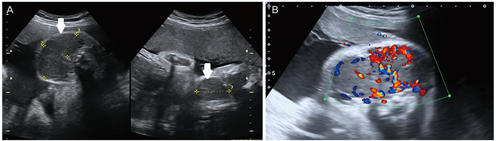
Fig. 1 (A) Two-dimensional ultrasound in 34th gestational week, on the axial and sagittal planes, showing the fetal goiter (arrows). (B) Color Doppler showing exuberant and uniform vascularization throughout the fetal thyroid.
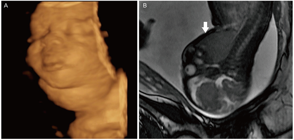
Fig. 2 Three-dimensional ultrasound in the rendering mode showing the fetal neck mass (A) and magnetic resonance image acquired in the sagittal view (B) showing the fetal goiter (arrow) in T2-weighted sequences.
Reference
-
1. Corbacioglu Esmer A, Gul A, Dagdeviren H, Turan Bakirci I, Sahin O. Intrauterine diagnosis and treatment of fetal goitrous hypothyroidism. J Obstet Gynaecol Res. 2013; 39:720–723.2. Pereira RC, Barroso LM, Mendes MJ, Joaquim IF, Ornelas H. A newborn with neck mass. Einstein. 2011; 9:78–80.3. Mayor-Lynn KA, Rohrs HJ 3rd, Cruz AC, Silverstein JH, Richards D. Antenatal diagnosis and treatment of a dyshormonogenetic fetal goiter. J Ultrasound Med. 2009; 28:67–71.4. Stewart CJ, Constantatos S, Joolay Y, Muller L. In utero treatment of fetal goitrous hypothyroidism in a euthyroid mother: a case report. J Clin Ultrasound. 2012; 40:603–606.5. Harreld JH, Kilani RK, Lascola CD, Bartz SK. MR imaging of fetal goiter. AJNR Am J Neuroradiol. 2011; 32:E160.6. Sanz-Cortes M, Fernandez S, Puerto B. Fetal thyroid masses and fetal goiter. In : Copel JA, D'Alton ME, Gratacos E, Platt LD, Tutschek B, Feltovich H, editors. Obstetric imaging. 1st ed. Philadelphia: Elsevier;2012. p. 378–386.7. Mirsky DM, Shekdar KV, Bilaniuk LT. Fetal MRI: head and neck. Magn Reson Imaging Clin N Am. 2012; 20:605–618.8. O'Connor SC, Rooks VJ, Smith AB. Magnetic resonance imaging of the fetal central nervous system, head, neck, and chest. Semin Ultrasound CT MR. 2012; 33:86–101.9. Baba K, Imanishi Y, Igarashi R, Shinagawa T. Evaluation of signal intensity of various thyroid tissues on MR imaging. St Marianna Med J. 2001; 29:303–311.10. Huel C, Guibourdenche J, Vuillard E, Ouahba J, Piketty M, Oury JF, et al. Use of ultrasound to distinguish between fetal hyperthyroidism and hypothyroidism on discovery of a goiter. Ultrasound Obstet Gynecol. 2009; 33:412–420.11. Jain V, Sharma R, Verma S, Agarwal R. Fetal euthyroid goiter. Indian J Pediatr. 2009; 76:1259–1260.12. Marin RC, Bello-Munoz JC, Martinez GV, Martinez SA, Moratonas EC, Roura LC. Use of 3-dimensional sonography for prenatal evaluation and follow-up of fetal goitrous hypothyroidism. J Ultrasound Med. 2010; 29:1339–1343.
- Full Text Links
-
- Actions
-
Cited
- CITED
-
- Close
- Share
-
- Similar articles
-
- Prenatal Diagnosis of Fetal Goiter in a Euthyroid Mother
- A Case of Prenatal Diagnosis of Congenital Fetal Goiter in Hyperthyroidism Mother
- Relationship of Prenatal Stress and Depression to Maternal-Fetal Attachment and Fetal Growth
- A Case of Fetal Cholelithiasis Related to Maternal Intrahepatic Cholestasis of Pregnancy
- Perinatal outcome of pregnancy with hyperthyroidism
