Cholesterol Granuloma of the Breast Incidentally Detected on Dynamic Abdominal CT: A Case Report
- Affiliations
-
- 1Department of Radiology, Soonchunhyang University College of Medicine, Bucheon Hospital, Bucheon, Korea. grace@schmc.ac.kr
- 2Department of Pathology, Soonchunhyang University College of Medicine, Bucheon Hospital, Bucheon, Korea.
- KMID: 2150459
- DOI: http://doi.org/10.3348/jksr.2016.74.1.22
Abstract
- A breast cholesterol granuloma is an uncommon nodular breast lesion. We incidentally detected a persistently enhancing breast mass on the dynamic abdominal computed tomography (CT) of a 78-year-old woman. The mass decreased in diameter over 50 days following a core needle biopsy. This report is the first to describe the dynamic-enhanced CT features of a breast cholesterol granuloma.
MeSH Terms
Figure
-
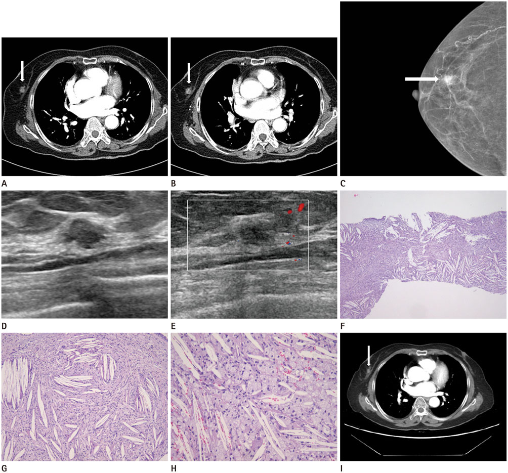
Fig. 1 Radiologic and pathologic findings of cholesterol granuloma in a 78-year-old woman. A. CT reveals a 1 cm diameter, oval, ill-defined, heterogeneously-enhanced mass in the right mid-outer breast (arrow). B. On delayed-enhanced CT, the enhancement is found to be progressive (arrow). C. A craniocaudal view of mammography reveals a 1 cm diameter, oval, indistinct hyperdense mass lacking microcalcification in the mid-outer portion of the right breast (arrow), corresponding to the enhanced mass evident on the dynamic abdominal CT. D, E. Ultrasonography of the right breast reveals an oval isoechoic mass with partially angular margins (D), but lacking increased intratumoral vascularity on color Doppler imaging (E). F-H. The cholesterol granuloma contains numerous cholesterol clefts and dense inflammatory infiltrations (H&E staining, × 40) (F), scatters cholesterol clefts surrounded by giant cells, histiocytes, and inflammatory cells (H&E staining, × 100) (G), and a dense infiltration of foamy histiocytes (H&E staining, × 200) (H). I. The diameter of the biopsy-proven cholesterol granuloma in the right breast has decreased by the time of the follow-up CT (arrow). H&E = hematoxylin-and-eosin
Reference
-
1. Wilhelmus JL, Schrodt GR, Mahaffey LM. Cholesterol granulomas of the breast. A lesion which clinically mimics carcinoma. Am J Clin Pathol. 1982; 77:592–597.2. Reynolds HE, Cramer HM. Cholesterol granuloma of the breast: a mimic of carcinoma. Radiology. 1994; 191:249–250.3. Osada T, Kitayama J, Nagawa H. Cholesterol granuloma of the breast mimicking carcinoma: report of a case. Surg Today. 2002; 32:981–984.4. Ahn HS, Kim SM, Yun BL, Kim MS, Jang M, Park SY, et al. The unusual ultrasound features of a breast cholesterol granuloma manifesting as an intracystic mass: case report and literature review. Korean J Radiol. 2013; 14:179–182.5. Bezić J, Piljić-Burazer M. Breast cholesterol granuloma: a report of two cases with discussion on potential pathogenesis. Pathologica. 2013; 105:349–352.6. Ishizaki M, Ohsumi S, Takashima S, Mandai K. Two cases of cholesterol granuloma of the breast. Breast Cancer. 2001; 8:158–161.7. Fujii T, Yajima R, Morita H, Yamaguchi S, Tsutsumi S, Asao T, et al. Cholesterol granuloma of the breast suspected as breast carcinoma. Int J Case Rep Images. 2013; 4:723–726.8. Döring L, Wedemeier G. [Cholesterol granuloma of the female breast]. Chirurg. 1974; 45:520–521.9. Fujita T, Doihara H, Takabatake D, Takahashi H, Yoshitomi S, Ishibe Y, et al. Multidetector row computed tomography for diagnosing intraductal extension of breast carcinoma. J Surg Oncol. 2005; 91:10–16.10. Sardanelli F, Calabrese M, Zandrino F, Melani E, Parodi R, Imperiale A, et al. Dynamic helical CT of breast tumors. J Comput Assist Tomogr. 1998; 22:398–407.
- Full Text Links
-
- Actions
-
Cited
- CITED
-
- Close
- Share
-
- Similar articles
-
- Cholesterol Granuloma of the Breast: A Case Report
- Incidentally detected laryngeal granuloma during orotracheal intubation under direct laryngoscope: A case report
- Cholesterol Granuloma Presenting as Retroperitoneal Mass: A case report
- A Case of Cholesterol Granuloma in Pancreas
- The Unusual Ultrasound Features of a Breast Cholesterol Granuloma Manifesting as an Intracystic Mass: Case Report and Literature Review
