The Unusual Ultrasound Features of a Breast Cholesterol Granuloma Manifesting as an Intracystic Mass: Case Report and Literature Review
- Affiliations
-
- 1Department of Radiology, Seoul National University Bundang Hospital, Seongnam 463-707, Korea. kimsmlms@paran.com
- 2Department of Pathology, Seoul National University Bundang Hospital, Seongnam 463-707, Korea.
- 3Department of Surgery, Seoul National University Bundang Hospital, Seongnam 463-707, Korea.
- 4Department of Radiology, Gyeongsang National University Hospital, Jinju 660-702, Korea.
- KMID: 1482775
- DOI: http://doi.org/10.3348/kjr.2013.14.2.179
Abstract
- Cholesterol granuloma of the breast is a rare, benign disease. Here, we present the unique ultrasonographic findings of breast cholesterol granuloma manifesting as an intracystic mass. The findings of this case report may help expand existing knowledge regarding differential diagnosis of intracystic breast masses, which are found on ultrasonographic examination.
Keyword
MeSH Terms
Figure
-
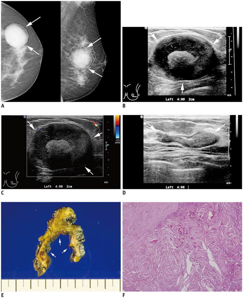
Fig. 1 Imaging findings of breast cholesterol granuloma in 62-year-old woman. A. Left mammography scan showing well-circumscribed, round high density mass (arrows). Mass exhibited coarse heterogeneous calcifications and peripheral radiolucency. B, C. Ultrasonography scan of left breast showing complex echoic mass (arrows), containing intracystic lobular, solid lesion (B) without vascularity on color Doppler imaging (C). There are no other abnormalities adjacent to mass. D. Core needle biopsy was performed for left breast mass. Mass collapsed after procedure (arrows), and approximately 20 mL of brownish fluid was aspirated. E, F. Gross and microscopic findings of cholesterol granuloma. 4.5 × 4.0 cm cystic lesion (arrows) is evident on cut section (E). Microscopically, characteristic needle-like cholesterol crystals and aggregates of histiocytes are found (F, hematoxylin and eosin, × 400).
Reference
-
1. Garofalo S, Casolino C, Accurso A, Falleti J. Cholesterol granuloma of the breast with unusual ossification features (osseous metaplasia). Pathol Res Pract. 2008. 204:353–356.2. Osada T, Kitayama J, Nagawa H. Cholesterol granuloma of the breast mimicking carcinoma: report of a case. Surg Today. 2002. 32:981–984.3. Furuhira C, Ohshima A, Shimada K, Kuroki S, Nakano K, Ishikawa M, et al. A case of breast cholesterol granuloma accompanied by cancer. Breast Cancer. 2004. 11:210–213.4. Kim JE, Park IS, Lee KH, Kim MY, Kim YJ. Cholesterol granuloma of the breast: a case report. J Korean Radiol Soc. 2008. 58:627–629.5. Smith GL, Hicks P, Wijesinghe DP, Holme TC. Cholesterol granuloma of the breast presenting as an intracystic papilloma. Br J Radiol. 1997. 70:1178–1179.6. Ganesan S, Karthik G, Joshi M, Damodaran V. Ultrasound spectrum in intraductal papillary neoplasms of breast. Br J Radiol. 2006. 79:843–849.
- Full Text Links
-
- Actions
-
Cited
- CITED
-
- Close
- Share
-
- Similar articles
-
- Cholesterol Granuloma of the Breast: A Case Report
- Cholesterol Granuloma of the Breast Incidentally Detected on Dynamic Abdominal CT: A Case Report
- Cholesterol Granuloma Presenting as Retroperitoneal Mass: A case report
- Fine Needle Aspiration Cytology of Intracystic Papillary Carcinoma of the Breast
- A Case of Huge Cholesterol Granuloma Cyst in Temporal Bone with Intracranial Extension
