J Korean Neurosurg Soc.
2013 Nov;54(5):420-422. 10.3340/jkns.2013.54.5.420.
Lymphomatosis Cerebri
- Affiliations
-
- 1Department of Neurosurgery, Ilsan Paik Hospital, College of Medicine, Inje University, Goyang, Korea. cychoi@paik.ac.kr
- 2Department of Pathology, Ilsan Paik Hospital, College of Medicine, Inje University, Goyang, Korea.
- KMID: 2138359
- DOI: http://doi.org/10.3340/jkns.2013.54.5.420
Abstract
- Lymphomatosis cerebri is considered a diffuse form of primary central nervous system lymphoma and very rare. It is not well recognized and may be misdiagnosed with infiltrating tumors, degenerative disorders, ischemic diseases, and infectious diseases developed in the brain. Awareness of the possibility of this rare disease and early biopsy are required for differential diagnosis and preventing poor clinical outcomes. We report a case with lymphomatosis cerebri who presented with rapid neurological deteriorations and review the relevant literatures.
MeSH Terms
Figure
-
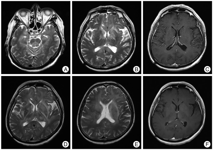
Fig. 1 MRI shows diffuse, poorly-circumscribed T2 hyper-intense lesions which involve the brainstem and deep regions in both cerebral hemispheres at presentation (A and B). These lesions are not enhanced with Gadolinium (C). Two months later, these lesions extend into both thalamus and periventricular deep white matters (D and E). Definite enhancement is still not noted (F).
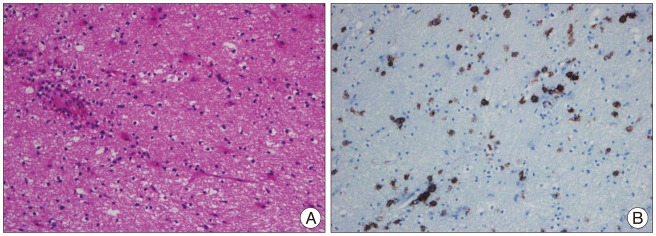
Fig. 2 H&E microphotograph displays scattered atypical lymphocytes and perivascular lymphoid cells (A). On immunohistochemical staining, atypical lymphoid cells are strongly stained with CD20 (B).
Reference
-
1. Abrey LE, Yahalom J, DeAngelis LM. Treatment for primary CNS lymphoma : the next step. J Clin Oncol. 2000; 18:3144–3150. PMID: 10963643.2. Ashworth B. Cerebral histiocytic lymphoma presenting with loss of weight. Neurology. 1982; 32:894–896. PMID: 7048129.
Article3. Chen S, Tanaka S, Giannini C, Morris J, Yan ES, Buckner J, et al. Gliomatosis cerebri : clinical characteristics, management, and outcomes. J Neurooncol. 2013; 112:267–275. PMID: 23341100.
Article4. Courtois F, Gille M, Haven F, Hantson P. Lymphomatosis cerebri presenting as a recurrent leukoencephalopathy. Case Rep Neurol. 2012; 4:181–186. PMID: 23185172.
Article6. Kanai R, Shibuya M, Hata T, Hori M, Hirabayashi K, Terada T, et al. A case of 'lymphomatosis cerebri' diagnosed in an early phase and treated by whole brain radiation : case report and literature review. J Neurooncol. 2008; 86:83–88. PMID: 17611716.
Article7. Keswani A, Bigio E, Grimm S. Lymphomatosis cerebri presenting with orthostatic hypotension, anorexia, and paraparesis. J Neurooncol. 2012; 109:581–586. PMID: 22806340.
Article8. Kitai R, Hashimoto N, Yamate K, Ikawa M, Yoneda M, Nakajima T, et al. Lymphomatosis cerebri : clinical characteristics, neuroimaging, and pathological findings. Brain Tumor Pathol. 2012; 29:47–53. PMID: 21927864.
Article9. Lewerenz J, Ding X, Matschke J, Schnabel C, Emami P, von Borczyskowski D, et al. Dementia and leukoencephalopathy due to lymphomatosis cerebri. J Neurol Neurosurg Psychiatry. 2007; 78:777–778. PMID: 17210623.
Article10. Raz E, Tinelli E, Antonelli M, Canevelli M, Fiorelli M, Bozzao L, et al. MRI findings in lymphomatosis cerebri : description of a case and revision of the literature. J Neuroimaging. 2011; 21:e183–e186. PMID: 20345746.
- Full Text Links
-
- Actions
-
Cited
- CITED
-
- Close
- Share
-
- Similar articles
-
- Evolution of Neurolymphomatosis to Lymphomatosis Cerebri
- A Case of Pseudotumor Cerebri Associated with Primary Antiphospholipid Syndrome
- The Ventriculoperitoneal Shunting in Pseudotumor Cerebri: Report of 2 Cases
- Computed tomographic findings of peritoneal lymphomatosis in a cat: a case report
- A Case of Pseudotumor Cerebri Associated with Systemic Lupus Erythematosus
