Differential Diagnosis of Inflammatory Bowel Disease: What Is the Role of Colonoscopy?
- Affiliations
-
- 1Department of Internal Medicine, Ewha Womans University School of Medicine, Seoul, Korea. jassa@ewha.ac.kr
Abstract
- Colonoscopy plays a crucial role in the diagnosis, treatment and follow-up monitoring of inflammatory bowel disease (IBD). Practitioners should be well informed of the colonoscopic findings of IBD to prevent the misdiagnosis, overtreatment or delayed treatment. Distinguishing between Crohn's disease and ulcerative colitis is essential in terms of pharmacological treatment, surgical decision-making, and prognosis. But there are still lesions with difficulty in differentiation that approximately 10% of the patients fall into the category of indeterminate colitis. Efforts are needed to carefully select treatment approach appropriate for each patient by providing a precise diagnosis on the extent and degree of lesions as well as to accurately delineate the lesions to assure that they are compared in subsequent rounds of follow-up monitoring in order to allow redetermination and adjustment of the treatment.
MeSH Terms
Figure
-
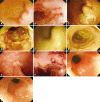
Fig. 1 Chronographic alteration in endoscopic findings of typical Crohn's disease. A 37-year-old female had colonoscopy with complaints of diarrhea. At initial diagnosis, multiple aphthous erosion and linear ulcer with normal surrounding mucosa were noted (A, B). She was diagnosed as Crohn's disease. After 2 years, colonoscopy revealed the ulcers were deeper with typical cobblestone appearance caused by numerous, confluent ulcerations (C, D). She had been treated with combination of steroid and immunosuppressants. Five months after the treatment, ulcers were healed with fibrotic change (E, F) with resolution of symptoms, and complete remission was achieved. Four years after the initial diagnosis, symptoms were exacerbated and follow-up colonoscopy showed deep ulcers with recurrent cobble stone appearance (G, H) that biologic agent was initiated. Even though the ulcerative lesions in the mucosa improved, stenosis followed, eventually leading to balloon dilation (I, J). Since the patient clearly showed natural progression during the treatment of Crohn's disease, there was no confusion in the diagnosis of Crohn's disease.
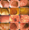
Fig. 2 Confusing endoscopic findings of ulcerative colitis. A 22-year-old male had colonoscopy with complaints of loose stool. At initial diagnosis, diffuse erythema with friable and granular mucosal change was noted from the rectum to the descending colon without involvement of the right colon (A, B, C). He was diagnosed as ulcerative colitis and had been treated for 1 year, and complete remission was achieved (D, E, F). Four years after the diagnosis, during the follow-up colonoscopy, multiple longitudinal ulcers without involvement of rectum were noted which could have been confused with Crohn's disese if the patient did not have colonoscopy previously (G, H, I). Colonoscopic findings were more compatible with ulcerative colitis 8 months later compared with the latest exam (J, K, L).
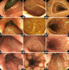
Fig. 3 Confusing endoscopic findings of Crohn's disease. A 22-year-old male had colonoscopy with complaints of mucoid stool. Initial findings showed friable mucosal change with granularity noted in the rectum without any involvement of the rest of the colon (A, B, C). The patient was diagnosed as ulcerative proctitis. One year later, disease was extended to the entire colon and the patient had systemic treatment with consideration of the extensive ulcerative colitis (D, E, F). Six months after the treatment, follow-up colonoscopy revealed that the rectum was spared with multiple linear skipped ulcer (G, H, I). Four years after the initial diagnosis, the rectum was involved again confusing the diagnosis. One year after the latest exam, cobblestone appearance was found (J, K, L). Final diagnosis was adjusted to Crohn's disease since he was initially diagnosed as ulcerative colitis 4 years ago
Reference
-
1. Yang SK, Byeon JS. Diagnosis and Treatment of Colonoscopy. 2009. Seoul: Koonja.2. Choi KY. Endoscopic manifestation of inflammatory bowel disease: ulcerative colitis. Proceedings of the 19th Seminar of Korean Society of Gastrointestinal Endoscopy. 1998. 1998 Aug 30-Sep 3; Seoul, Korea. Seoul: Korean Society of Gastrointestinal Endoscopy;p. 47–54.3. Yang SK. Endoscopic diagnosis of ulcerative colitis. Proceedings of the 22nd Seminar of Korean Society of Gastrointestinal Endoscopy. 2000. 2003 Mar 26; Seoul, Korea. Seoul: Korean Society of Gastrointestinal Endoscopy;p. 201–218.4. Kim TI. Endoscopic differentiation of inflammatory lesions: IBD. Korean J Gastrointest Endosc. 2003; 27:119–125.5. Chutkan RK, Wayne JD. Kirsner JB, editor. Endoscopy in inflammatory bowel disease. Inflammatory Bowel Disease. 2000. 5th ed. Baltimore: Williams and Wilkins;p. 453–477.6. Bernstein CN, Shanahan F, Anton PA, Weinstein WM. Patchiness of mucosal inflammation in treated ulcerative colitis: a prospective study. Gastrointest Endosc. 1995; 42:232–237. PMID: 7498688.
Article7. Matsumoto T, Nakamura S, Shimizu M, Iida M. Significance of appendiceal involvement in patients with ulcerative colitis. Gastrointest Endosc. 2002; 55:180–185. PMID: 11818919.
Article8. Yang SK, Jung HY, Kang GH, et al. Appendiceal orifice inflammation as a skip lesion in ulcerative colitis: an analysis in relation to medical therapy and disease extent. Gastrointest Endosc. 1999; 49:743–747. PMID: 10343220.
Article9. Deutsch DE, Olson AD. Colonoscopy or sigmoidoscopy as the initial evaluation of pediatric patients with colitis: a survey of physician behavior and a cost analysis. J Pediatr Gastroenterol Nutr. 1997; 25:26–31. PMID: 9226523.
Article10. Kim WH. Endoscopic manifestation of inflammatory bowel disease: Crohn's disease. Proceedings of the 19th Seminar of Korean Society of Gastrointestinal Endoscopy. 1998. 1998 Aug 30-Sep 3; Seoul, Korea. Seoul: Korean Society of Gastrointestinal Endoscopy;p. 55–67.11. Kim HJ. Crohn's disease. Proceedings of the 22nd Seminar of Korean Society of Gastrointestinal Endoscopy. 2000. 2003 Mar 26; Seoul, Korea. Seoul: Korean Society of Gastrointestinal Endoscopy;p. 209–215.12. Fujimura Y, Kamoi R, Iida M. Pathogenesis of aphthoid ulcers in Crohn's disease: correlative findings by magnifying colonoscopy, electron microscopy, and immunohistochemistry. Gut. 1996; 38:724–732. PMID: 8707119.
Article13. Ramzan NN, Leighton JA, Heigh RI, Shapiro MS. Clinical significance of granuloma in Crohn's disease. Inflamm Bowel Dis. 2002; 8:168–173. PMID: 11979136.
Article14. Korelitz BI, Sommers SC. Rectal biopsy in patients with Crohn's disease. Normal mucosa on sigmoidoscopic examination. JAMA. 1977; 237:2742–2744. PMID: 577228.
Article15. Herrerías JM, Caunedo A, Rodríguez-Téllez M, Pellicer F, Herrerías JM Jr. Capsule endoscopy in patients with suspected Crohn's disease and negative endoscopy. Endoscopy. 2003; 35:564–568. PMID: 12822090.16. Kim YH. Common errors in the diagnosis process. Intest Res. 2003; 1:106–110.17. Pera A, Bellando P, Caldera D, et al. Colonoscopy in inflammatory bowel disease. Diagnostic accuracy and proposal of an endoscopic score. Gastroenterology. 1987; 92:181–185. PMID: 3781186.18. Moum B, Ekbom A, Vatn MH, et al. Clinical course during the 1st year after diagnosis in ulcerative colitis and Crohn's disease. Results of a large, prospective population-based study in southeastern Norway, 1990-93. Scand J Gastroenterol. 1997; 32:1005–1012. PMID: 9361173.
Article19. Alcántara M, Rodriguez R, Potenciano JL, Carrobles JL, Muñoz C, Gomez R. Endoscopic and bioptic findings in the upper gastrointestinal tract in patients with Crohn's disease. Endoscopy. 1993; 25:282–286. PMID: 8330547.
Article20. Lo SK, Zaidel O, Tabibzadeh S, et al. Utility of wireless capsule endoscopy and IBD serology in re-classifying indeterminatecolitis. Gastroenterology. 2003; 124:s1310.21. Tedesco FJ, Hardin RD, Harper RN, Edwards BH. Infectious colitis endoscopically simulating inflammatory bowel disease: a prospective evaluation. Gastrointest Endosc. 1983; 29:195–197. PMID: 6618115.
Article22. Fochios SE, Korelitz BI. The role of sigmoidoscopy and rectal biopsy in diagnosis and management of inflammatory bowel disease: personal experience. Am J Gastroenterol. 1988; 83:114–119. PMID: 3341332.23. Matsumoto T, Iida M, Kimura Y, Fujishima M. Culture of colonoscopically obtained biopsy specimens in acute infectious colitis. Gastrointest Endosc. 1994; 40(2 Pt 1):184–187. PMID: 8013819.
Article24. Surawicz CM, Belic L. Rectal biopsy helps to distinguish acute self-limited colitis from idiopathic inflammatory bowel disease. Gastroenterology. 1984; 86:104–113. PMID: 6689653.
Article25. Meucci G, Vecchi M, Astegiano M, et al. The natural history of ulcerative proctitis: a multicenter, retrospective study. Gruppo di Studio per le Malattie Infiammatorie Intestinali (GSMII). Am J Gastroenterol. 2000; 95:469–473. PMID: 10685752.26. Langholz E, Munkholm P, Davidsen M, Nielsen OH, Binder V. Changes in extent of ulcerative colitis: a study on the course and prognostic factors. Scand J Gastroenterol. 1996; 31:260–266. PMID: 8833356.
Article27. Baron JH, Connell AM, Lennard-Jones JE. Variation between observers in describing mucosal appearances in proctocolitis. Br Med J. 1964; 1:89–92. PMID: 14075156.
Article28. Alemayehu G, Järnerot G. Colonoscopy during an attack of severe ulcerative colitis is a safe procedure and of great value in clinical decision making. Am J Gastroenterol. 1991; 86:187–190. PMID: 1992632.29. Modigliani R, Mary JY, Simon JF, et al. Clinical, biological, and endoscopic picture of attacks of Crohn's disease. Evolution on prednisolone Groupe d'Etude. Thérapeutique des Affections Inflammatoires Digestives. Gastroenterology. 1990; 98:811–818. PMID: 2179031.30. Hanauer SB, Feagan BG, Lichtenstein GR, et al. Maintenance infliximab for Crohn's disease: the ACCENT I randomised trial. Lancet. 2002; 359:1541–1549. PMID: 12047962.
Article31. Pineton de Chambrun G, Peyrin-Biroulet L, Lémann M, Colombel JF. Clinical implications of mucosal healing for the management of IBD. Nat Rev Gastroenterol Hepatol. 2010; 7:15–29. PMID: 19949430.
Article32. Sipponen T, Nuutinen H, Turunen U, Färkkilä M. Endoscopic evaluation of Crohn's disease activity: comparison of the CDEIS and the SES-CD. Inflamm Bowel Dis. 2010; 16:2131–2136. PMID: 20848462.33. Lichtenstein GR, Rutgeerts P. Importance of mucosal healing in ulcerative colitis. Inflamm Bowel Dis. 2010; 16:338–346. PMID: 19637362.
Article34. Travis SP, Schnell D, Krzeski P, et al. Developing an instrument to assess the endoscopic severity of ulcerative colitis: the Ulcerative Colitis Endoscopic Index of Severity (UCEIS). Gut. 2012; 61:535–542. PMID: 21997563.
Article35. Truelove SC, Witts LJ. Cortisone in ulcerative colitis; final report on a therapeutic trial. Br Med J. 1955; 2:1041–1048. PMID: 13260656.
- Full Text Links
-
- Actions
-
Cited
- CITED
-
- Close
- Share
-
- Similar articles
-
- Role of colonoscopy in the diagnosis and treatment of pediatric lower gastrointestinal disorders
- Imaging Techniques and Differential Diagnosis for Inflammatory Bowel Disease
- The Role of Colonoscopy in Inflammatory Bowel Disease
- A Case of Intestinal Tuberculosis Diagnosed by Colonoscopy
- Probe-based confocal laser endomicroscopy in the differential diagnosis of inflammatory bowel diseases: a case series
