Probe-based confocal laser endomicroscopy in the differential diagnosis of inflammatory bowel diseases: a case series
- Affiliations
-
- 1Department of Internal Medicine and Institute of Gastroenterology, Yonsei University College of Medicine, Seoul, Korea. geniushee@yuhs.ac
- KMID: 2434167
- DOI: http://doi.org/10.5217/ir.2018.00035
Abstract
- No abstract available.
Figure
-
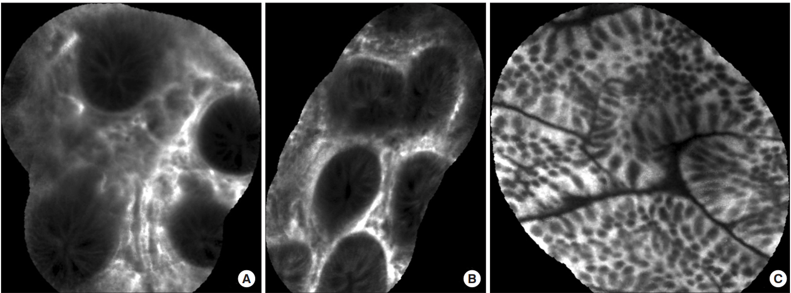
Fig. 1. Changes of the intestinal mucosa detected by probe-based confocal laser endomicroscopy in UC (Patient A). (A) Irregular architecture and increased distance between the colonic crypts. (B) Leakage of fluorescein into the crypt lumen from the interstitium of the colon. (C) Normal colonic crypts with no fluorescein in the lumen and no crypt distortion.

Fig. 2. Changes of the intestinal mucosa detected by probe-based confocal laser endomicroscopy in a CD (Patient C). (A, B) Ileocecal valve ulcer; crypt destruction, fluorescent spillage, no granuloma visible. (C) Straight appearance of crypts with luminal opening of the crypts resembling as black holes in the normal mucosa of sigmoid colon.

Fig. 3. Changes of the intestinal mucosa detected by probe-based confocal laser endomicroscopy in intestinal Behçet’s disease (Patient E). (A) Anastomosis site ulcer; crypt destruction with disarrayed feature. (B) Abundant inflammatory cell infiltration. Granuloma, AFB, or vasculitis not visible. (C) Normal colonic crypts with no fluorescein in the lumen and no crypt distortion.
Reference
-
1. Kiesslich R, Goetz M, Vieth M, Galle PR, Neurath MF. Confocal laser endomicroscopy. Gastrointest Endosc Clin N Am. 2005; 15:715–731.
Article2. Cheon JH, Kim ES, Shin SJ, et al. Development and validation of novel diagnostic criteria for intestinal Behçet’s disease in Korean patients with ileocolonic ulcers. Am J Gastroenterol. 2009; 104:2492–2499.
Article3. Dignass A, Eliakim R, Magro F, et al. Second European evidence-based consensus on the diagnosis and management of ulcerative colitis. Part 1: definitions and diagnosis (Spanish version). Rev Gastroenterol Mex. 2014; 79:263–289.4. Van Assche G, Dignass A, Panes J, et al. The second European evidence-based consensus on the diagnosis and management of Crohn’s disease: definitions and diagnosis. J Crohns Colitis. 2010; 4:7–27.
Article5. Chapman CG, Konda VJ. Confocal laser endomicroscopy in inflammatory bowel disease: achieving new depths in mucosal healing. Gastrointest Endosc. 2016; 83:792–794.
Article6. Cheon JH. Advances in the endoscopic assessment of inflammatory bowel diseases: cooperation between endoscopic and pathologic evaluations. J Pathol Transl Med. 2015; 49:209–217.
Article7. Cheon JH, Kim WH. Recent advances of endoscopy in inflammatory bowel diseases. Gut Liver. 2007; 1:118–125.
Article8. Rispo A, Castiglione F, Staibano S, et al. Diagnostic accuracy of confocal laser endomicroscopy in diagnosing dysplasia in patients affected by long-standing ulcerative colitis. World J Gastrointest Endosc. 2012; 4:414–420.
Article9. Vermeire S, Van Assche G, Rutgeerts P. Classification of inflammatory bowel disease: the old and the new. Curr Opin Gastroenterol. 2012; 28:321–326.10. Mowat C, Cole A, Windsor A, et al. Guidelines for the management of inflammatory bowel disease in adults. Gut. 2011; 60:571–607.
Article
- Full Text Links
-
- Actions
-
Cited
- CITED
-
- Close
- Share
-
- Similar articles
-
- High-Resolution Probe-Based Confocal Laser Endomicroscopy for Diagnosing Biliary Diseases
- Role of Advanced Endoscopic Imaging Techniques in the Management of Inflammatory Bowel Disease
- A Review of Probe-Based Confocal Laser Endomicroscopy for Pancreaticobiliary Disease
- Application and Efficacy of Super-Magnifying Endoscopy for the Lower Intestinal Tract
- Confocal Laser Endomicroscopy and Molecular Imaging in Barrett Esophagus and Stomach
