Yonsei Med J.
2014 Jul;55(4):944-952. 10.3349/ymj.2014.55.4.944.
Optical Coherence Tomographic Observation of Morphological Features of Neointimal Tissue after Drug-Eluting Stent Implantation
- Affiliations
-
- 1Division of Cardiology, Severance Cardiovascular Hospital, Yonsei University College of Medicine, Seoul, Korea. mkhong61@yuhs.ac
- 2Severance Biomedical Science Institute, Yonsei University College of Medicine, Seoul, Korea.
- KMID: 2130818
- DOI: http://doi.org/10.3349/ymj.2014.55.4.944
Abstract
- PURPOSE
The impacts of different time courses and the degree of neointimal growth on neointimal morphology have not yet been sufficiently investigated. Therefore, we evaluated the morphological features of neointimal tissue after drug-eluting stent (DES) implantation using optical coherence tomography (OCT).
MATERIALS AND METHODS
The morphological features of neointimal tissue in stented segments with a maximal percentage of cross-sectional area (CSA) stenosis of neointima were evaluated in 507 DES-treated lesions with >100 microm mean neointimal thickness on follow-up OCT. Neointimal tissue was categorized as homogeneous, heterogeneous, layered, or neoatherosclerotic.
RESULTS
In lesions with <50% of neointimal CSA stenosis, homogeneous neointima (68.2%) was predominant, followed by heterogeneous neointima (14.1%) and layered neointima (14.1%). In lesions with > or =50% of neointimal CSA stenosis, layered neointima was most frequently observed (68.3%), followed by neoatherosclerotic neointima (25.2%). In subgroup analysis of lesions with > or =50% of neointimal CSA stenosis, 89.5% of the lesions with a stent age <30 months were layered neointima, while 62.3% of the lesions with a stent age > or =30 months were neoatherosclerotic neointima.
CONCLUSION
This study suggests that the OCT-detected morphology of DES neointimal tissue was different according to the follow-up time course and degree of neointimal hyperplasia.
MeSH Terms
Figure
-
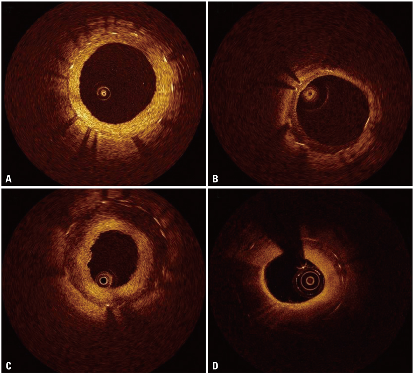
Fig. 1 Representative optical coherence tomographic images of neointimal tissue. (A) Homogeneous type. (B) Heterogeneous type. (C) Layered type. (D) Neoatherosclerotic type.
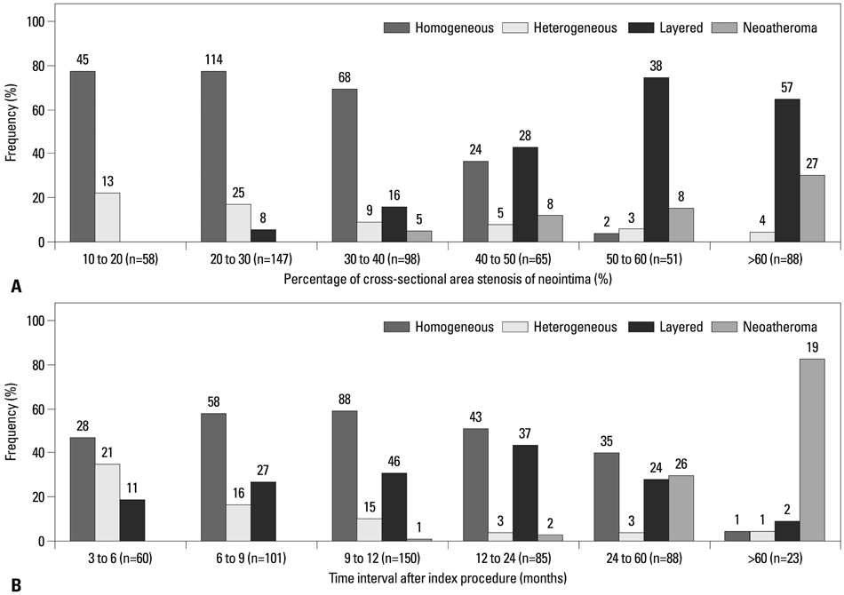
Fig. 2 Distribution of various neointimal tissues according to the percentage of neointimal cross-sectional area stenosis (A) and time interval of optical coherence tomography after index procedure (B).
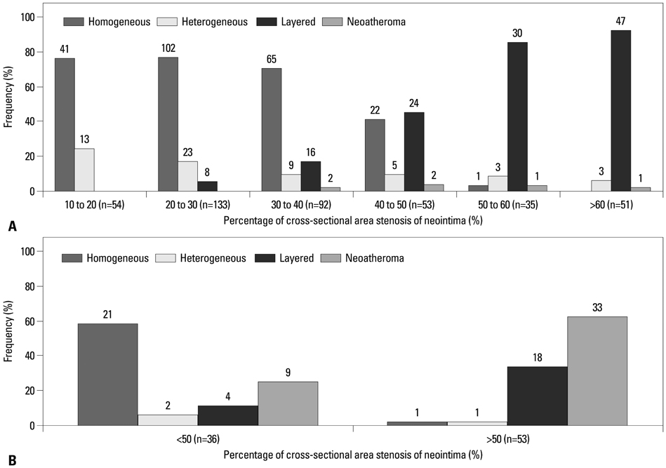
Fig. 3 Distribution of various neointimal tissues according to the percentage of neointimal cross-sectional area stenosis in lesions of stent age <30 months (A) and ≥30 months (B).
Reference
-
1. Gonzalo N, Serruys PW, Okamura T, van Beusekom HM, Garcia-Garcia HM, van Soest G, et al. Optical coherence tomography patterns of stent restenosis. Am Heart J. 2009; 158:284–293.
Article2. Lee SY, Hong MK. Stent evaluation with optical coherence tomography. Yonsei Med J. 2013; 54:1075–1083.
Article3. Kang SJ, Mintz GS, Akasaka T, Park DW, Lee JY, Kim WJ, et al. Optical coherence tomographic analysis of in-stent neoatherosclerosis after drug-eluting stent implantation. Circulation. 2011; 123:2954–2963.
Article4. Habara M, Terashima M, Nasu K, Kaneda H, Yokota D, Ito T, et al. Morphological differences of tissue characteristics between early, late, and very late restenosis lesions after first generation drug-eluting stent implantation: an optical coherence tomography study. Eur Heart J Cardiovasc Imaging. 2013; 14:276–284.
Article5. Lee SJ, Kim BK, Kim JS, Ko YG, Choi D, Jang Y, et al. Evaluation of neointimal morphology of lesions with or without in-stent restenosis: an optical coherence tomography study. Clin Cardiol. 2011; 34:633–639.
Article6. Yonetsu T, Kim JS, Kato K, Kim SJ, Xing L, Yeh RW, et al. Comparison of incidence and time course of neoatherosclerosis between bare metal stents and drug-eluting stents using optical coherence tomography. Am J Cardiol. 2012; 110:933–939.
Article7. Takano M, Yamamoto M, Inami S, Murakami D, Ohba T, Seino Y, et al. Appearance of lipid-laden intima and neovascularization after implantation of bare-metal stents extended late-phase observation by intracoronary optical coherence tomography. J Am Coll Cardiol. 2009; 55:26–32.
Article8. Kim U, Kim JS, Kim JS, Lee JM, Son JW, Kim J, et al. The initial extent of malapposition in ST-elevation myocardial infarction treated with drug-eluting stent: the usefulness of optical coherence tomography. Yonsei Med J. 2010; 51:332–338.
Article9. Prati F, Regar E, Mintz GS, Arbustini E, Di Mario C, Jang IK, et al. Expert review document on methodology, terminology, and clinical applications of optical coherence tomography: physical principles, methodology of image acquisition, and clinical application for assessment of coronary arteries and atherosclerosis. Eur Heart J. 2010; 31:401–415.
Article10. Lee SY, Shin DH, Mintz GS, Kim JS, Kim BK, Ko YG, et al. Optical coherence tomography-based evaluation of in-stent neoatherosclerosis in lesions with more than 50% neointimal cross-sectional area stenosis. EuroIntervention. 2013; 9:945–951.
Article11. Farb A, Sangiorgi G, Carter AJ, Walley VM, Edwards WD, Schwartz RS, et al. Pathology of acute and chronic coronary stenting in humans. Circulation. 1999; 99:44–52.
Article12. van Beusekom HM, Saia F, Zindler JD, Lemos PA, Swager-Ten Hoor SL, van Leeuwen MA, et al. Drug-eluting stents show delayed healing: paclitaxel more pronounced than sirolimus. Eur Heart J. 2007; 28:974–979.
Article13. Chieffo A, Foglieni C, Nodari RL, Briguori C, Sangiorgi G, Latib A, et al. Histopathology of clinical coronary restenosis in drug-eluting versus bare metal stents. Am J Cardiol. 2009; 104:1660–1667.
Article14. Nakano M, Vorpahl M, Otsuka F, Taniwaki M, Yazdani SK, Finn AV, et al. Ex vivo assessment of vascular response to coronary stents by optical frequency domain imaging. JACC Cardiovasc Imaging. 2012; 5:71–82.
Article15. Nagai H, Ishibashi-Ueda H, Fujii K. Histology of highly echolucent regions in optical coherence tomography images from two patients with sirolimus-eluting stent restenosis. Catheter Cardiovasc Interv. 2010; 75:961–963.
Article16. Yonetsu T, Kato K, Kim SJ, Xing L, Jia H, McNulty I, et al. Predictors for neoatherosclerosis: a retrospective observational study from the optical coherence tomography registry. Circ Cardiovasc Imaging. 2012; 5:660–666.
- Full Text Links
-
- Actions
-
Cited
- CITED
-
- Close
- Share
-
- Similar articles
-
- A Newly Formed and Ruptured Atheromatous Plaque within Neointima after Drug-Eluting Stent Implantation: 2-Year Follow-Up Intravascular Ultrasound and Optical Coherence Tomography Studies
- Clinical Implication of Optical Coherence Tomography-Based Neoatherosclerosis
- Late Stent Thrombosis After Drug-Eluting Stent Implantation: A Rare Case of Accelerated Neo-Atherosclerosis and Early Manifestation of Neointimal Rupture
- Formation and Transformation of Neointima after Drug-eluting Stent Implantation: Insights from Optical Coherence Tomographic Studies
- Comparison of Optical Coherence Tomographic Assessment between First- and Second-Generation Drug-Eluting Stents
