A Newly Formed and Ruptured Atheromatous Plaque within Neointima after Drug-Eluting Stent Implantation: 2-Year Follow-Up Intravascular Ultrasound and Optical Coherence Tomography Studies
- Affiliations
-
- 1Division of Cardiology, Yonsei Cardiovascular Center, Yonsei University College of Medicine, Seoul, Korea. mkhong61@yuhs.ac
- KMID: 1058827
- DOI: http://doi.org/10.3349/ymj.2011.52.6.1028
Abstract
- Late stent thrombosis (LST) which is a life threatening complication has emerged as a serious problem of drug-eluting stents (DES). Several studies have suggested that incomplete neointimal coverage of stent struts contributes to LST. Progressive atherosclerosis within the neointima is an another possible cause of LST, but this phenomenon has seldom been reported in DES. We present a case of LST following DES implantation after a period of 28 months due to ruptured atheromatous plaque, despite complete neointimal coverage of stent struts proven by optical coherence tomography.
MeSH Terms
Figure
-
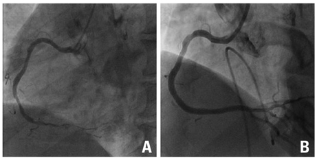
Fig. 1 Initial coronary angiogram of the RCA. (A) Coronary angiogram shows significant luminal narrowing at the distal RCA. (B) After stent implantation, there is a minimal residual stenosis. RCA, right coronary artery.
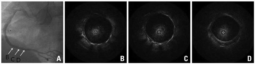
Fig. 2 Coronary angiogram and OCT findings at 12-month follow-up. (A)Coronary angiogram shows no restenosis in the stented segment. (B, C and D)The OCT shows complete neointimal coverage of stent struts. OCT, optical coherence tomography.

Fig. 3 (A) The 2nd follow-up angiogram at 28 months after stent implantation shows abnormal contrast filling in a previously implanted stent (see arrow). (B, C and D) IVUS and OCT images reveal newly formed and ruptured atheromatous plaque within neointima. Arrowheads indicate stent strut. IVUS , intravascular ultrasound; OCT, optical coherence tomography.
Reference
-
1. Iakovou I, Schmidt T, Bonizzoni E, Ge L, Sangiorgi GM, Stankovic G, et al. Incidence, predictors, and outcome of thrombosis after successful implantation of drug-eluting stents. JAMA. 2005. 293:2126–2130.
Article2. Lüscher TF, Steffel J, Eberli FR, Joner M, Nakazawa G, Tanner FC, et al. Drug-eluting stent and coronary thrombosis: biological mechanisms and clinical implications. Circulation. 2007. 115:1051–1058.3. Kim U, Kim JS, Kim JS, Lee JM, Son JW, Kim J, et al. The initial extent of malapposition in ST-elevation myocardial infarction treated with drug-eluting stent: the usefulness of optical coherence tomography. Yonsei Med J. 2010. 51:332–338.
Article4. Kotani J, Awata M, Nanto S, Uematsu M, Oshima F, Minamiguchi H, et al. Incomplete neointimal coverage of sirolimus-eluting stents: angioscopic findings. J Am Coll Cardiol. 2006. 47:2108–2111.5. Matsumoto D, Shite J, Shinke T, Otake H, Tanino Y, Ogasawara D, et al. Neointimal coverage of sirolimus-eluting stents at 6-month follow-up: evaluated by optical coherence tomography. Eur Heart J. 2007. 28:961–967.
Article6. Finn AV, Joner M, Nakazawa G, Kolodgie F, Newell J, John MC, et al. Pathological correlates of late drug-eluting stent thrombosis: strut coverage as a marker of endothelialization. Circulation. 2007. 115:2435–2441.
Article7. Higo T, Ueda Y, Oyabu J, Okada K, Nishio M, Hirata A, et al. Atherosclerotic and thrombogenic neointima formed over sirolimus drug-eluting stent: an angioscopic study. JACC Cardiovasc Imaging. 2009. 2:616–624.8. Doyle B, Rihal CS, O'Sullivan CJ, Lennon RJ, Wiste HJ, Bell M, et al. Outcomes of stent thrombosis and restenosis during extended follow-up of patients treated with bare-metal coronary stents. Circulation. 2007. 116:2391–2398.9. Nakazawa G, Vorpahl M, Finn AV, Narula J, Virmani R. One step forward and two steps back with drug-eluting-stents: from preventing restenosis to causing late thrombosis and nouveau atherosclerosis. JACC Cardiovasc Imaging. 2009. 2:625–628.
- Full Text Links
-
- Actions
-
Cited
- CITED
-
- Close
- Share
-
- Similar articles
-
- Formation and Transformation of Neointima after Drug-eluting Stent Implantation: Insights from Optical Coherence Tomographic Studies
- Optical Coherence Tomographic Observation of Morphological Features of Neointimal Tissue after Drug-Eluting Stent Implantation
- A Case of In-Stent Neointimal Plaque Rupture 10 Years After Bare Metal Stent Implantation: Intravascular Ultrasound and Optical Coherence Tomographic Findings
- Stent Evaluation with Optical Coherence Tomography
- Late Stent Thrombosis After Drug-Eluting Stent Implantation: A Rare Case of Accelerated Neo-Atherosclerosis and Early Manifestation of Neointimal Rupture
