CD34 and p53 Immunohistochemical Stains Differentiate Hypocellular Myelodysplastic Syndrome (hMDS) from Aplastic Anemia and a CD34 Immunohistochemical Stain Provides Useful Survival Information for hMDS
- Affiliations
-
- 1Department of Laboratory Medicine, University of Ulsan College of Medicine and Asan Medical Center, Seoul, Korea. cjpark@amc.seoul.kr, cchoongh@gnah.co.kr
- 2Department of Internal Medicine, University of Ulsan College of Medicine and Asan Medical Center, Seoul, Korea.
- 3Department of Pediatrics, University of Ulsan College of Medicine and Asan Medical Center, Seoul, Korea.
- 4Department of Laboratory Medicine, University of Ulsan College of Medicine and Gangneung Asan Hospital, Gangneung, Korea.
- KMID: 2129567
- DOI: http://doi.org/10.3343/alm.2014.34.6.426
Abstract
- BACKGROUND
The presence of significant dysplasia in bone marrow (BM) aspirates helps to distinguish between hypocellular myelodysplastic syndrome (hMDS) and aplastic anemia (AA). Occasionally, diluted BM aspirates make it difficult to recognize dysplastic changes and can also negatively affect the detection of cytogenetic abnormalities in hMDS. We evaluated the usefulness of CD34 and p53 immunoreactivity for discriminating between hMDS and AA and for estimating survival outcomes in hMDS patients.
METHODS
BM clot section (BMC) or BM biopsy (BMB) specimens were obtained from 64 hMDS/AA patients (33 with hMDS and 31 with AA) and seven controls. Immunohistochemical (IHC) staining for CD34 and p53 was performed by using the EnVision detection system (Dako, Denmark). We compared the results of IHC staining, BM findings, and chromosomal analyses, and determined overall survival outcomes.
RESULTS
The number of CD34- and p53-positive BM cells was higher among the patients with hMDS than among the patients with AA (P<0.001 and P=0.001, respectively). hMDS patients with increased CD34-positive cells had significantly poorer survival outcomes compared with those with normal number of CD34-positive cells (P=0.013).
CONCLUSIONS
CD34 and p53 IHC stains of BMC or BMB provide useful information for differentiating between hMDS and AA. CD34 IHC staining of BMC or BMB also provides useful information for estimating survival outcomes in hMDS patients.
MeSH Terms
-
Adolescent
Adult
Anemia, Aplastic/*diagnosis
Antigens, CD34/*metabolism
Bone Marrow/metabolism/*pathology
Child
Chromosome Aberrations
Diagnosis, Differential
Female
Humans
Immunohistochemistry
Kaplan-Meier Estimate
Male
Middle Aged
Myelodysplastic Syndromes/*diagnosis/mortality
ROC Curve
Tumor Suppressor Protein p53/*metabolism
Antigens, CD34
Tumor Suppressor Protein p53
Figure
-
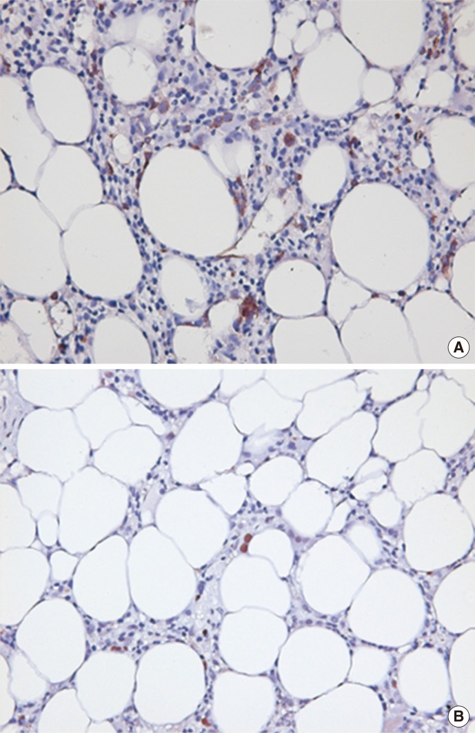
Fig. 1 Example of CD34+ (A) and p53+ (B) immunohistochemical stains in a patient with hypocellular myelodysplastic syndrome (bone marrow biopsy; immunohistochemical stain; original magnification ×400).

Fig. 2 Kaplan-Meier analysis comparing overall survival between the patients with hypocellular myelodysplastic syndrome (hMDS; N=33) and those with aplastic anemia (AA; N=31).
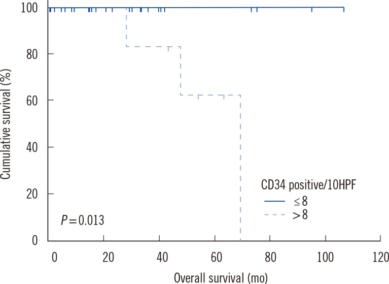
Fig. 3 Kaplan-Meier analysis for the overall survival of the patients with hypocellular myelodysplastic syndrome. The patients were divided into groups based on the CD34 immunohistochemistry status.
Cited by 1 articles
-
Clinical Relevance of p53 Immunohistochemical Stain in the Differential Diagnosis Between Pediatric Aplastic Anemia and Refractory Cytopenia of Childhood
Sang Hyuk Park, Hyun-Sook Chi, Young-Uk Cho, Seongsoo Jang, Chan-Jeoung Park, Ho-Joon Im, Jong-Jin Seo
Ann Lab Med. 2016;36(2):174-176. doi: 10.3343/alm.2016.36.2.174.
Reference
-
1. Orazi A, Albitar M, Heerema NA, Haskins S, Neiman RS. Hypoplastic myelodysplastic syndromes can be distinguished from acquired aplastic anemia by CD34 and PCNA immunostaining of bone marrow biopsy specimens. Am J Clin Pathol. 1997; 107:268–274. PMID: 9052376.
Article2. Orazi A, Czader MB. Myelodysplastic syndromes. Am J Clin Pathol. 2009; 132:290–305. PMID: 19605823.
Article3. Tuzuner N, Cox C, Rowe JM, Watrous D, Bennett JM. Hypocellular myelodysplastic syndromes (MDS): new proposals. Br J Haematol. 1995; 91:612–617. PMID: 8555063.
Article4. Baur AS, Meugé-Moraw C, Schmidt PM, Parlier V, Jotterand M, Delacrétaz F. CD34/QBEND10 immunostaining in bone marrow biopsies: an additional parameter for the diagnosis and classification of myelodysplastic syndromes. Eur J Haematol. 2000; 64:71–79. PMID: 10997326.5. Elghetany MT, Vyas S, Yuoh G. Significance of p53 overexpression in bone marrow biopsies from patients with bone marrow failure: aplastic anemia, hypocellular refractory anemia, and hypercellular refractory anemia. Ann Hematol. 1998; 77:261–264. PMID: 9875662.
Article6. Totzke G, Brüning T, Vetter H, Schulze-Osthoff K, Ko Y. P53 downregulation in myelodysplastic syndrome - a quantitative analysis by competitive RT-PCR. Leukemia. 2001; 15:1663–1664. PMID: 11587227.
Article7. Lepelley P, Preudhomme C, Vanrumbeke M, Quesnel B, Cosson A, Fenaux P. Detection of p53 mutations in hematological malignancies: comparison between immunocytochemistry and DNA analysis. Leukemia. 1994; 8:1342–1349. PMID: 8057671.8. Brunning RD, Orazi A, Germing U, Le Beau MM, Porwit A, Baumann I, et al. Myelodysplastic syndromes/neoplasms, overview. In : Swerdlow SH, Campo E, editors. WHO classification of tumours of haematopoietic and lymphoid tissues. 4th ed. Lyon: IARC;2008. p. 88–93.9. Greenberg PL, Tuechler H, Schanz J, Sanz G, Garcia-Manero G, Solé F, et al. Revised international prognostic scoring system for myelodysplastic syndromes. Blood. 2012; 120:2454–2465. PMID: 22740453.
Article10. Bain BJ, Clark DM, Wilkins BS. Acute myeloid leukemia, mixed phenotype acute leukemia, the myelodysplastic syndromes and histiocytic neoplasms. In : Bain BJ, Clark DM, editors. Bone marrow pathology. 4th ed. Oxford: Wiley-Blackwell;2010. p. 166–238.11. Bain BJ. The myelodysplastic syndromes. In : Bain BJ, editor. Leukaemia diagnosis. 4th ed. Oxford: Wiley-Blackwell;2010. p. 219–260.12. Brodsky RA. Acquired aplastic anemia. In : Greer JP, Foerster J, editors. Wintrobe's clinical hematology. 12th ed. Philadelphia: Lippincott Williams & Wilkins;2009. p. 1185–1195.13. Matsui WH, Brodsky RA, Smith BD, Borowitz MJ, Jones RJ. Quantitative analysis of bone marrow CD34 cells in aplastic anemia and hypoplastic myelodysplastic syndromes. Leukemia. 2006; 20:458–462. PMID: 16437138.
Article14. Choi JW, Fujino M, Ito M. F-blast is a useful marker for differentiating hypocellular refractory anemia from aplastic anemia. Int J Hematol. 2002; 75:257–260. PMID: 11999352.
Article15. Elghetany MT. p53 overexpression in bone marrow biopsies in refractory anemia and aplastic anemia: impact of antibody selection. Leuk Res. 2000; 24:975–977. PMID: 11086182.
Article16. Horny HP, Sotlar K, Valent P. Diagnostic value of histology and immunohistochemistry in myelodysplastic syndromes. Leuk Res. 2007; 31:1609–1616. PMID: 17604834.
Article17. De Raeve H, Van Marck E, Van Camp B, Vanderkerken K. Angiogenesis and the role of bone marrow endothelial cells in haematological malignancies. Histol Histopathol. 2004; 19:935–950. PMID: 15168356.
- Full Text Links
-
- Actions
-
Cited
- CITED
-
- Close
- Share
-
- Similar articles
-
- Tissue Microarray Analysis of the Expression of p53, c-kit and CD34 in Sarcomas
- Successful engraftment after infusion of multiple low doses of CD34+ cells from a poorly matched sibling donor in a patient with severe aplastic anemia
- Clinical Relevance of p53 Immunohistochemical Stain in the Differential Diagnosis Between Pediatric Aplastic Anemia and Refractory Cytopenia of Childhood
- CD34 immunohistochemical staining of bone marrow biopsies in myelodysplastic syndromes
- Successful Selective CD34+ Cell Infusion after Late Graft Failure of Hematopoietic Stem Cell Transplantation in Two Cases of Severe Aplastic Anemia
