Intramuscular Hemangioma in the Anterior Scalene Muscle Diagnosed by Core Needle Biopsy
- Affiliations
-
- 1Department of Otorhinolaryngology-Head and Neck Surgery, Pusan National University Hospital, Busan, Korea. Cha.Wonjae@gmail.com
- 2Biomedical Research Institute, Pusan National University Hospital, Busan, Korea.
- 3Department of Otorhinolaryngology-Head and Neck Surgery, Seoul National University Hospital, Seoul, Korea.
- KMID: 2117528
- DOI: http://doi.org/10.3342/ceo.2015.8.3.298
Abstract
- Intramuscular hemangioma (IMH) is a rare, benign vascular lesion that frequently develops within skeletal muscles. Preoperatively, accurate diagnosis of IMH is often extremely difficult because of nonspecific clinical findings and the inaccuracy of fine-needle aspiration cytology. IMH is suspected in only 8% of preoperative diagnoses before surgical exploration. Here, we report a case of a 44-year-old man with a huge IMH in the anterior scalene muscle that was preoperatively diagnosed using ultrasonography-guided core needle biopsy, and was successfully treated based on preoperative clinical information.
MeSH Terms
Figure
-
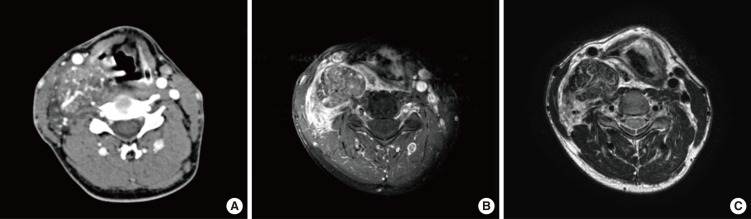
Fig. 1 Neck computed tomography (CT) revealed a hypervascular mass, approximately 8.5 cm in size, at the right posterior cervical space (A). On CT, the right carotid artery was anteriorly displaced, and the mass showed isosignal intensity relative to muscle. Magnetic resonance imaging (MRI) was diagnostic and revealed a well-defined mass with slightly increased signal intensity on gadolinium-enhanced T1-weighted fat-saturated imaging (B) and slight increased signal intensity on T2-weighted imaging (C) within the right anterior scalene muscle.
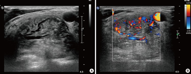
Fig. 2 Ultrasonography (US) showed a hypoechogenic and hypervascular mass involving the right parapharyngeal space posterolateral to the right common carotid artery. US-guided core needle biopsy was performed without complications.
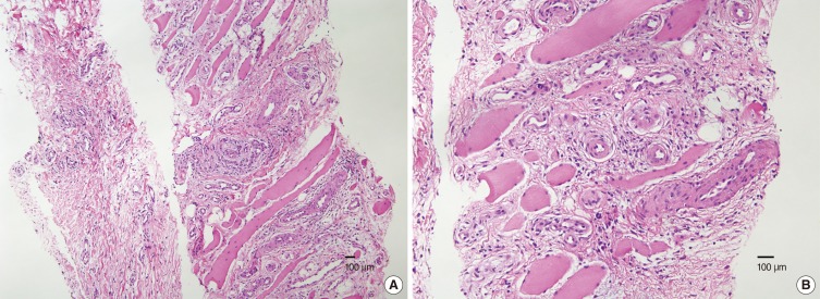
Fig. 3 The core needle biopsy specimen revealed skeletal muscle fragments with increased vasculature and adipose tissue that was diagnosed as intramuscular hemangioma (H&E: A, ×100; B, ×200).
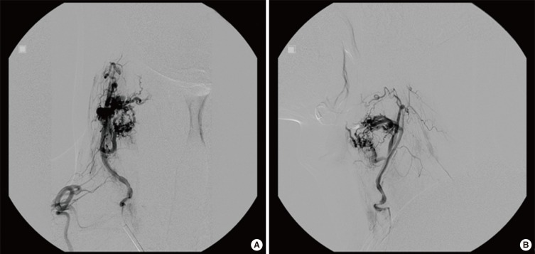
Fig. 4 Angiography and tumor embolization were planned 3 days before the elective operation. Angiography demonstrated a hypervascular mass fed by a branch of the right thyrocervical artery, and the mass was successfully embolized. (A) Anterior to posterior view, (B) lateral view.
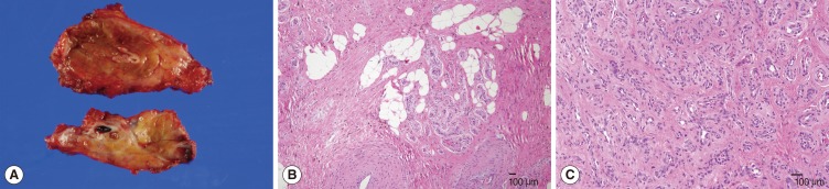
Fig. 5 (A) The specimen revealed increased vascularity among the muscle fibers. The histopathologic diagnosis was consistent with intramuscular hemangioma (H&E: B, ×100; C, ×200).
Reference
-
1. Giudice M, Piazza C, Bolzoni A, Peretti G. Head and neck intramuscular haemangioma: report of two cases with unusual localization. Eur Arch Otorhinolaryngol. 2003; 10. 260(9):498–501. PMID: 12748867.
Article2. Chaudhary N, Jain A, Gudwani S, Kapoor R, Motwani G. Intramuscular haemangioma of head and neck region. J Laryngol Otol. 1998; 12. 112(12):1199–1201. PMID: 10209624.
Article3. Okabe Y, Ishikawa S, Furukawa M. Intramuscular hemangioma of the masseter muscle: its characteristic appearance on magnetic resonance imaging. ORL J Otorhinolaryngol Relat Spec. 1991; 53(6):366–369. PMID: 1784478.
Article4. Welsh D, Hengerer AS. The diagnosis and treatment of intramuscular hemangiomas of the masseter muscle. Am J Otolaryngol. 1980; 2. 1(2):186–190. PMID: 7446838.
Article5. Scott JE. Haemangiomata in skeletal muscle. Br J Surg. 1957; 3. 44(187):496–501. PMID: 13510618.6. Ferlito A, Nicolai P, Gale N. Intramuscular haemangioma of the middle scalene muscle. Acta Otorhinolaryngol Belg. 1980; 34(3):345–349. PMID: 7234376.7. Van Abel KM, Carlson ML, Janus JR, Torres-Mora J, Moore EJ, Olsen KD, et al. Intramuscular hemangioma of the scalene musculature masquerading as a paraganglioma: a case series. Am J Otolaryngol. 2013; Mar-Apr. 34(2):158–162. PMID: 23159015.
Article8. Itoh K, Nishimura K, Togashi K, Fujisawa I, Nakano Y, Itoh H, et al. MR imaging of cavernous hemangioma of the face and neck. J Comput Assist Tomogr. 1986; Sep-Oct. 10(5):831–835. PMID: 3745555.
Article9. Liston R. Case of erectile tumour in the popliteal space.-removal. Med Chir Trans. 1843; 26:120–132.
Article10. Salzman R, Buchanan MA, Berman L, Jani P. Ultrasound-guided core-needle biopsy and magnetic resonance imaging in the accurate diagnosis of intramuscular haemangiomas of the head and neck. J Laryngol Otol. 2012; 4. 126(4):391–394. PMID: 22258504.
Article11. Moumoulidis I, Durvasula VS, Jani P. An unusual neck lump: intramuscular haemangioma of the sternocleidomastoid muscle. Eur Arch Otorhinolaryngol. 2007; 10. 264(10):1257–1260. PMID: 17593381.
Article12. Stofman GM, Reiter D, Feldman MD. Invasive intramuscular hemangiomas of the head and neck. Ear Nose Throat J. 1989; 8. 68(8):612–616. PMID: 2583030.
- Full Text Links
-
- Actions
-
Cited
- CITED
-
- Close
- Share
-
- Similar articles
-
- Cavernous Hemangioma of the Masseter Muscle
- Intramuscular Hemangioma of the Mentalis Muscle: A Case Report
- Intramuscular hemangioma formation in the masseter muscle: a case report
- Pseudoaneurysm of the Breast after Core Needle Biopsy: A Case Report
- A Case of Intramuscular Muller Muscle Hemangioma of Upper Eyelid Mimicking Sarcoidosis
