Intramuscular hemangioma formation in the masseter muscle: a case report
- Affiliations
-
- 1Department of Oral and Maxillofacial Surgery, College of dentistry, Yonsei University, Seoul, Korea. Kimoms@yuhs.ac
Abstract
- Hemangioma is a benign vascular proliferation. Intramuscular hemangiomas are rare, accounting for less than 1% of all hemangiomas, and occur normally in the trunk and extremities. Approximately 10-20% of intramuscular hemangiomas are found in the head and neck region, most often in the masseter muscles. The typical clinical characteristic is a painful soft tissue mass without cutaneous changes. The suggested treatment is a surgical excision. We report a case of an intramuscular hemagnioma of the masseter muscle. The patient was a 56 year old male who visited our clinic complaining of left facial swelling after 2 years of follow up at a different clinic. After magnetic resonance imaging (MRI), the mass was excised under general anesthesia. The biopsy revealed the mass to be an intramuscular hemangioma. We report the clinical and pathological characteristics as well as the treatment of a case of an intramuscular hemangioma of the masseter muscle.
MeSH Terms
Figure
-

Fig. 1. Preoperative magnetic resonance imaging (MRI) shows 3.3 cm size, soft issue mass at Lt. masticatory space. The mass shows intermediate signal on T2WI and strong enhancement. It is embedded in anterior side of Lt. masseter muscle with no bony invasion. A is the T1 weighted MRI, B is the enhanced T1 weighted MRI, and C is the T2 weighted MRI.
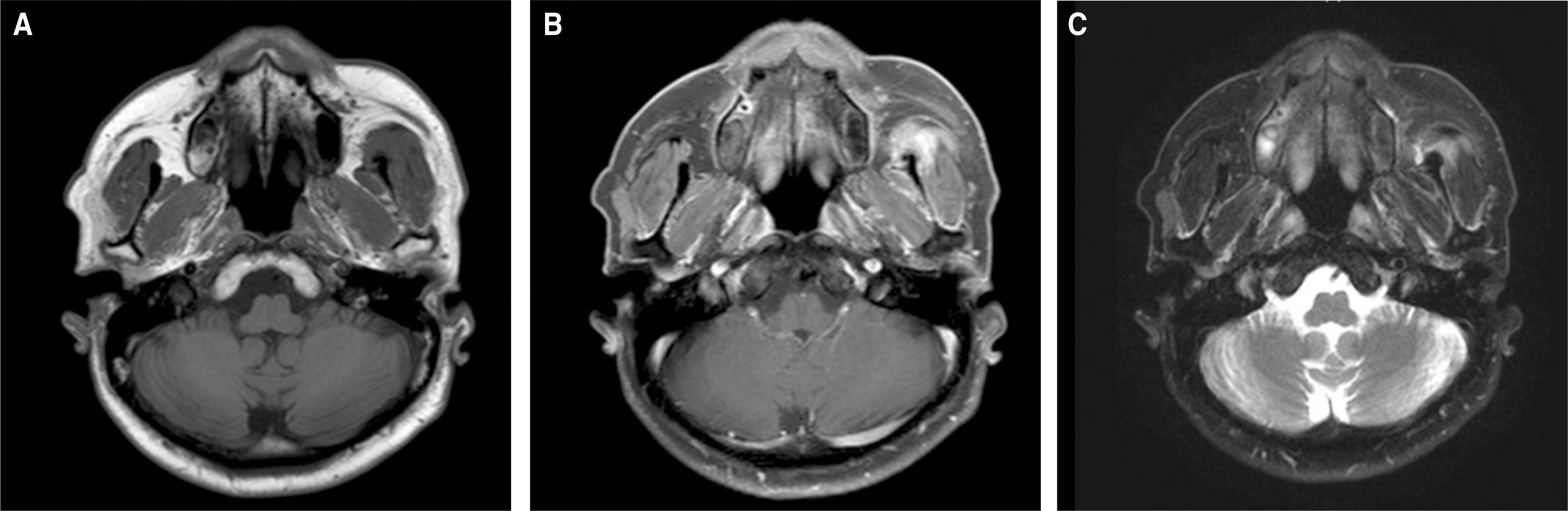
Fig. 2. Magnetic resonance imaging (MRI) of postoperative 3 months shows small residual lesion on anterior position of Lt. masseter muscle. A is the T1 weighted MRI, B is the enhanced T1 weighted MRI, and C is the T2 weighted MRI.
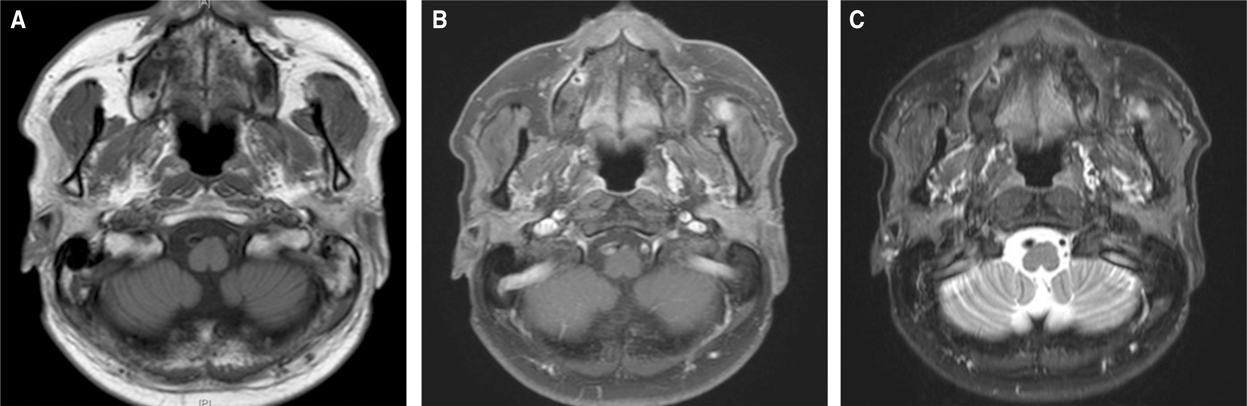
Fig. 3. Magnetic resonance imaging (MRI) of postoperative 12 months shows residual lesion on Lt. masticator space with localizing and decreasing in size. A is the T1 weighted MRI, B is the enhanced T1 weighted MRI, and C is the T2 weighted MRI.
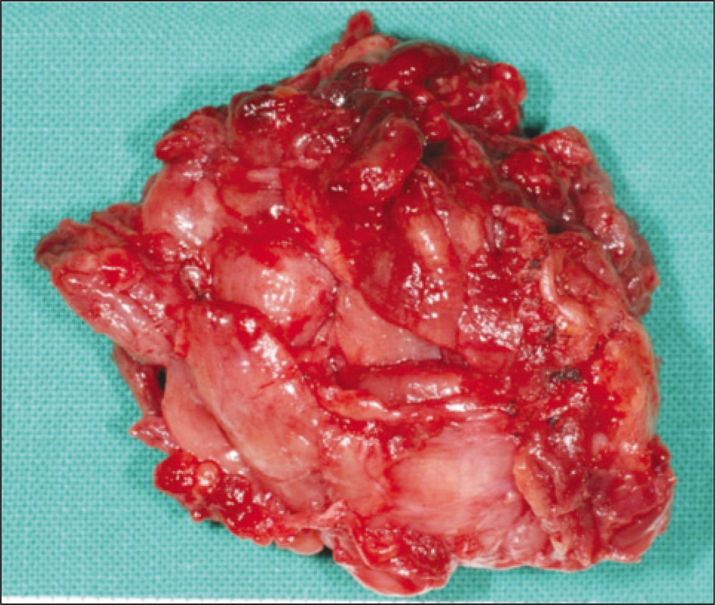
Fig. 4. Main mass is measured 4.5×3.5×4.0 cm.

Fig. 5. A. Microscopic examination demonstrates multiple muscles (H & E staining, original magnificationx 40), B. Microscopic examination demonstrates many vessels inside muscles (H & E staining, original magnificationx 200).
Reference
-
References
1. Sato M, Shirasuna K, Imai J, Miyazaki T. Giant cavernous hemangioma of the masseter muscle: report of case. J Oral Surg. 1979; 37:496–9.2. Shallow TA, Eger SA, Wagner FB. Primary hemangiomatous tumors of skeletal muscle. Ann Surg. 1944; 119:700–40.
Article3. Lee SK, Kwon SY. Intramuscular cavernous hemangioma arising from masseter muscle: a diagnostic dilemma (2006: 12b). Eur Radiol. 2007; 17:854–7.
Article4. Wolf GT, Daniel F, Krause CJ, Kaufman RS. Intramuscular hemangioma of the head and neck. Laryngoscope. 1985; 95:210–3.
Article5. Narayanan CD, Prakash P, Dhanasekaran CK. Intramuscular hemangioma of the masseter muscle: a case report. Cases J. 2009; 2:7459.
Article6. Ingalls GK, Bonnington GJ, Sisk AL. Intramuscular hemangioma of the mentalis muscle. Oral Surg Oral Med Oral Pathol. 1985; 60:476–81.
Article7. Rossiter JL, Hendrix RA, Tom LW, Potsic WP. Intramuscular hemangioma of the head and neck. Otolaryngol Head Neck Surg. 1993; 108:18–26.
Article8. Hoehn JG, Farrow GM, Devine KD, Masson JK. Invasive hemangioma of the head and neck. Am J Surg. 1970; 120:495–500.
Article9. Persky MS, Berenstein A, Cohen NL. Combined treatment of head and neck vascular masses with preoperative embolization. Laryngoscope. 1984; 94:20–7.
Article10. Sayan NB, Kogo M, Koizumi H, Watatani K, Saka M, Matsuya T, et al. Intramuscular hemangioma in the digastric muscle. J Osaka Univ Dent Sch. 1992; 32:14–20.11. Elahi MM, Parnes L, Fox A. Hemangioma of the masseter muscle. J Otolaryngol. 1992; 21:177–9.12. Rogalski R, Hensinger R, Loder R. Vascular abnormalities of the extremities: clinical findings and management. J Pediatr Orthop. 1993; 13:9–14.13. Morris SJ, Adams H. Case report: paediatric intramuscular hae-mangiomata-don' t overlook the phlebolith! Br J Radiol. 1995; 68:208–11.14. Odabasi AO, Metin KK, Mutlu C, Bas¸ak S, Erpek G. Intramuscular hemangioma of the masseter muscle. Eur Arch Otorhinolaryngol. 1999; 256:366–9.
Article15. Buetow PC, Kransdorf MJ, Moser RP Jr, Jelinek JS, Berrey BH. Radiologic appearance of intramuscular hemangioma with emphasis on MR imaging. AJR Am J Roentgenol. 1990; 154:563–7.
Article16. Kim KW, Sang JK, Cheong JH. Sclerotherapy of benign oral vascular lesion with sodium tetradecyl sulfate: cases report. J Korean Assoc Oral Maxillofac Surg. 2010; 36:280–5.
Article
- Full Text Links
-
- Actions
-
Cited
- CITED
-
- Close
- Share
-
- Similar articles
-
- Cavernous Hemangioma of the Masseter Muscle
- A hemangioma in the masseter muscle: a case report
- Intramuscular Hemangioma of the Sternocleidomastoid Muscle: An Unusual Neck Mass
- Intramuscular Hemangioma of the Mentalis Muscle: A Case Report
- Intramuscular hemangioma in the zygomaticus minor muscle: a case report and literature review
