J Korean Neurosurg Soc.
2015 Oct;58(4):357-362. 10.3340/jkns.2015.58.4.357.
The Effects of Spinopelvic Parameters and Paraspinal Muscle Degeneration on S1 Screw Loosening
- Affiliations
-
- 1Department of Neurosurgery, Chung-Ang University Hospital, Seoul, Korea. nspsw@cau.ac.kr
- 2Department of Neurosurgery, Gangneung Asan Hospital, University of Ulsan College of Medicine, Gangneung, Korea.
- KMID: 2114373
- DOI: http://doi.org/10.3340/jkns.2015.58.4.357
Abstract
OBJECTIVE
To investigate risk factors for S1 screw loosening after lumbosacral fusion, including spinopelvic parameters and paraspinal muscles.
METHODS
We studied with 156 patients with degenerative lumbar disease who underwent lumbosacral interbody fusion and pedicle screw fixation including the level of L5-S1 between 2005 and 2012. The patients were divided into loosening and non-loosening groups. Screw loosening was defined as a halo sign larger than 1 mm around a screw. We checked cross sectional area of paraspinal muscles, mean signal intensity of the muscles on T2 weight MRI as a degree of fatty degeneration, spinopelvic parameters, bone mineral density, number of fusion level, and the characteristic of S1 screw.
RESULTS
Twenty seven patients showed S1 screw loosening, which is 24.4% of total. The mean duration for S1 screw loosening was 7.3+/-4.1 months after surgery. Statistically significant risk factors were increased age, poor BMD, 3 or more fusion levels (p<0.05). Among spinopelvic parameters, a high pelvic incidence (p<0.01), a greater difference between pelvic incidence and lumbar lordotic angle preoperatively (p<0.01) and postoperatively (p<0.05). Smaller cross-sectional area and high T2 signal intensity in both multifidus and erector spinae muscles were also significant muscular risk factors (p<0.05). Small converging angle (p<0.001) and short intraosseous length (p<0.05) of S1 screw were significant screw related risk factors (p<0.05).
CONCLUSION
In addition to well known risk factors, spinopelvic parameters and the degeneration of paraspinal muscles also showed significant effects on the S1 screw loosening.
MeSH Terms
Figure
-

Fig. 1 Halo sign around S1 screws (arrows) on simple radiograph (A) and CT (B).
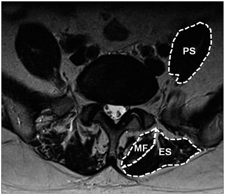
Fig. 2 Paraspinal muscles at L5-S1 level, multifidus muscle (MF), erector spinae muscle (ES), and psoas muscle (PS).
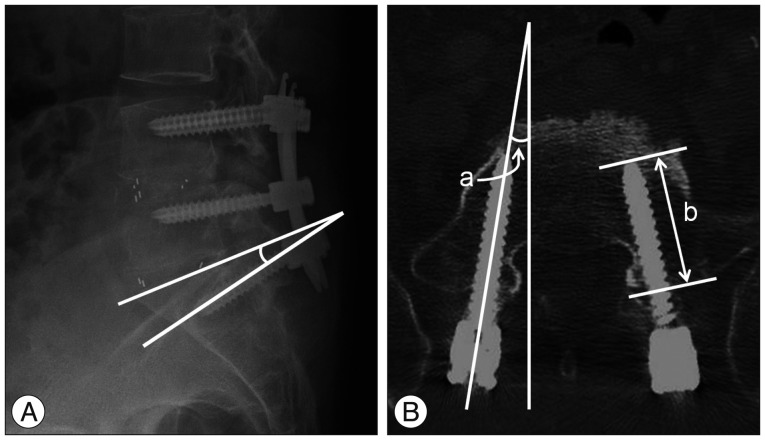
Fig. 3 A : Sagittal angle of S1 screw between screw and upper end plate of S1 vertebra on simple lateral radiograph. B : Axial angle of S1 screw between screw and vertical line (a) and intraosseous length of S1 screw as the length of intraosseous portion of the screw on postoperative CT (b).
Reference
-
1. Allen BL Jr, Ferguson RL. The Galveston technique of pelvic fixation with L-rod instrumentation of the spine. Spine (Phila Pa 1976). 1984; 9:388–394. PMID: 6474253.
Article2. Balderston RA, Winter RB, Moe JH, Bradford DS, Lonstein JE. Fusion to the sacrum for nonparalytic scoliosis in the adult. Spine (Phila Pa 1976). 1986; 11:824–829. PMID: 3027899.
Article3. Barber JW, Boden SD, Ganey T, Hutton WC. Biomechanical study of lumbar pedicle screws : does convergence affect axial pullout strength? J Spinal Disord. 1998; 11:215–220. PMID: 9657546.4. Bernhardt M, Swartz DE, Clothiaux PL, Crowell RR, White AA 3rd. Posterolateral lumbar and lumbosacral fusion with and without pedicle screw internal fixation. Clin Orthop Relat Res. 1992; (284):109–115. PMID: 1395279.
Article5. Cho W, Mason JR, Smith JS, Shimer AL, Wilson AS, Shaffrey CI, et al. Failure of lumbopelvic fixation after long construct fusions in patients with adult spinal deformity : clinical and radiographic risk factors : clinical article. J Neurosurg Spine. 2013; 19:445–453. PMID: 23909551.
Article6. Dakhil-Jerew F, Jadeja H, Cohen A, Shepperd JA. Inter-observer reliability of detecting Dynesys pedicle screw using plain X-rays : a study on 50 post-operative patients. Eur Spine J. 2009; 18:1486–1493. PMID: 19533178.
Article7. Dekutoski MB, Schendel MJ, Ogilvie JW, Olsewski JM, Wallace LJ, Lewis JL. Comparison of in vivo and in vitro adjacent segment motion after lumbar fusion. Spine (Phila Pa 1976). 1994; 19:1745–1751. PMID: 7973970.
Article8. Devlin VJ, Boachie-Adjei O, Bradford DS, Ogilvie JW, Transfeldt EE. Treatment of adult spinal deformity with fusion to the sacrum using CD instrumentation. J Spinal Disord. 1991; 4:1–14. PMID: 1839666.9. Emami A, Deviren V, Berven S, Smith JA, Hu SS, Bradford DS. Outcome and complications of long fusions to the sacrum in adult spine deformity : luque-galveston, combined iliac and sacral screws, and sacral fixation. Spine (Phila Pa 1976). 2002; 27:776–786. PMID: 11923673.
Article10. Finger T, Bayerl S, Onken J, Czabanka M, Woitzik J, Vajkoczy P. Sacropelvic fixation versus fusion to the sacrum for spondylodesis in multilevel degenerative spine disease. Eur Spine J. 2014; 23:1013–1020. PMID: 24448893.
Article11. Goutallier D, Postel JM, Bernageau J, Lavau L, Voisin MC. Fatty muscle degeneration in cuff ruptures. Pre- and postoperative evaluation by CT scan. Clin Orthop Relat Res. 1994; (304):78–83. PMID: 8020238.12. Herkowitz HN. Lumbar spinal stenosis : indications for arthrodesis and spinal instrumentation. Instr Course Lect. 1994; 43:425–433. PMID: 9097172.13. Huang Y, Majumdar S, Genant HK, Chan WP, Sharma KR, Yu P, et al. Quantitative MR relaxometry study of muscle composition and function in Duchenne muscular dystrophy. J Magn Reson Imaging. 1994; 4:59–64. PMID: 8148557.
Article14. Humphreys SC, Hodges SD, Patwardhan AG, Eck JC, Murphy RB, Covington LA. Comparison of posterior and transforaminal approaches to lumbar interbody fusion. Spine (Phila Pa 1976). 2001; 26:567–571. PMID: 11242386.
Article15. Jorgensen MJ, Marras WS, Gupta P. Cross-sectional area of the lumbar back muscles as a function of torso flexion. Clin Biomech (Bristol, Avon). 2003; 18:280–286.
Article16. Kim EH, Kim HJ. Long segment fusion to L5 vertebra and sacral vertebra in degenerative lumbar spine. J Korean Soc Spine Surg. 2002; 9:216–222.
Article17. Kim HK, Laor T, Horn PS, Racadio JM, Wong B, Dardzinski BJ. T2 mapping in Duchenne muscular dystrophy : distribution of disease activity and correlation with clinical assessments. Radiology. 2010; 255:899–908. PMID: 20501727.
Article18. Kim YJ, Bridwell KH, Lenke LG, Cho KJ, Edwards CC 2nd, Rinella AS. Pseudarthrosis in adult spinal deformity following multisegmental instrumentation and arthrodesis. J Bone Joint Surg Am. 2006; 88:721–728. PMID: 16595461.
Article19. Kim YJ, Bridwell KH, Lenke LG, Rhim S, Cheh G. Pseudarthrosis in long adult spinal deformity instrumentation and fusion to the sacrum : prevalence and risk factor analysis of 144 cases. Spine (Phila Pa 1976). 2006; 31:2329–2336. PMID: 16985461.
Article20. Kostuik JP, Hall BB. Spinal fusions to the sacrum in adults with scoliosis. Spine (Phila Pa 1976). 1983; 8:489–500. PMID: 6648699.
Article21. Krag MH, Beynnon BD, Pope MH, Frymoyer JW, Haugh LD, Weaver DL. An internal fixator for posterior application to short segments of the thoracic, lumbar, or lumbosacral spine. Design and testing. Clin Orthop Relat Res. 1986; (203):75–98. PMID: 3956000.
Article22. La Grone MO. Loss of lumbar lordosis. A complication of spinal fusion for scoliosis. Orthop Clin North Am. 1988; 19:383–393. PMID: 3282206.
Article23. Lazennec JY, Ramaré S, Arafati N, Laudet CG, Gorin M, Roger B, et al. Sagittal alignment in lumbosacral fusion : relations between radiological parameters and pain. Eur Spine J. 2000; 9:47–55. PMID: 10766077.
Article24. Lebwohl NH, Cunningham BW, Dmitriev A, Shimamoto N, Gooch L, Devlin V, et al. Biomechanical comparison of lumbosacral fixation techniques in a calf spine model. Spine (Phila Pa 1976). 2002; 27:2312–2320. PMID: 12438978.
Article25. Lee CS, Chung SS, Choi SW, Yu JW, Sohn MS. Critical length of fusion requiring additional fixation to prevent nonunion of the lumbosacral junction. Spine (Phila Pa 1976). 2010; 35:E206–E211. PMID: 20195201.
Article26. Lee JC, Cha JG, Kim Y, Kim YI, Shin BJ. Quantitative analysis of back muscle degeneration in the patients with the degenerative lumbar flat back using a digital image analysis : comparison with the normal controls. Spine (Phila Pa 1976). 2008; 33:318–325. PMID: 18303466.
Article27. Lehman RA Jr, Kuklo TR, Belmont PJ Jr, Andersen RC, Polly DW Jr. Advantage of pedicle screw fixation directed into the apex of the sacral promontory over bicortical fixation : a biomechanical analysis. Spine (Phila Pa 1976). 2002; 27:806–811. PMID: 11935101.
Article28. Leong JC, Lu WW, Zheng Y, Zhu Q, Zhong S. Comparison of the strengths of lumbosacral fixation achieved with techniques using one and two triangulated sacral screws. Spine (Phila Pa 1976). 1998; 23:2289–2294. PMID: 9820908.
Article29. Luk KD, Chen L, Lu WW. A stronger bicortical sacral pedicle screw fixation through the s1 endplate : an in vitro cyclic loading and pull-out force evaluation. Spine (Phila Pa 1976). 2005; 30:525–529. PMID: 15738784.
Article30. McCarthy RE, Bruffett WL, McCullough FL. S rod fixation to the sacrum in patients with neuromuscular spinal deformities. Clin Orthop Relat Res. 1999; (364):26–31. PMID: 10416388.
Article31. McLachlin SD, Al Saleh K, Gurr KR, Bailey SI, Bailey CS, Dunning CE. Comparative assessment of sacral screw loosening augmented with PMMA versus a calcium triglyceride bone cement. Spine (Phila Pa 1976). 2011; 36:E699–E704. PMID: 21289585.
Article32. Mengiardi B, Schmid MR, Boos N, Pfirrmann CW, Brunner F, Elfering A, et al. Fat content of lumbar paraspinal muscles in patients with chronic low back pain and in asymptomatic volunteers : quantification with MR spectroscopy. Radiology. 2006; 240:786–792. PMID: 16926328.
Article33. Moore DC, Maitra RS, Farjo LA, Graziano GP, Goldstein SA. Restoration of pedicle screw fixation with an in situ setting calcium phosphate cement. Spine (Phila Pa 1976). 1997; 22:1696–1705. PMID: 9259778.
Article34. Pihlajämaki H, Myllynen P, Böstman O. Complications of transpedicular lumbosacral fixation for non-traumatic disorders. J Bone Joint Surg Br. 1997; 79:183–189. PMID: 9119839.35. Rechtine GR, Sutterlin CE, Wood GW, Boyd RJ, Mansfield FL. The efficacy of pedicle screw/plate fixation on lumbar/lumbosacral autogenous bone graft fusion in adult patients with degenerative spondylolisthesis. J Spinal Disord. 1996; 9:382–391.
Article36. Renner SM, Lim TH, Kim WJ, Katolik L, An HS, Andersson GB. Augmentation of pedicle screw fixation strength using an injectable calcium phosphate cement as a function of injection timing and method. Spine (Phila Pa 1976). 2004; 29:E212–E216. PMID: 15167670.
Article37. Roy-Camille R, Saillant G, Mazel C. Internal fixation of the lumbar spine with pedicle screw plating. Clin Orthop Relat Res. 1986; (203):7–17. PMID: 3955999.
Article38. Ruland CM, McAfee PC, Warden KE, Cunningham BW. Triangulation of pedicular instrumentation. A biomechanical analysis. Spine (Phila Pa 1976). 1991; 16(6 Suppl):S270–S276.39. Sandén B, Olerud C, Petrén-Mallmin M, Johansson C, Larsson S. The significance of radiolucent zones surrounding pedicle screws. Definition of screw loosening in spinal instrumentation. J Bone Joint Surg Br. 2004; 86:457–461.40. Schwab F, Lafage V, Patel A, Farcy JP. Sagittal plane considerations and the pelvis in the adult patient. Spine (Phila Pa 1976). 2009; 34:1828–1833. PMID: 19644334.
Article41. Seo JB, Yoo JS, Jang HS, Kim JS. Correlation of clinical symptoms and function with fatty degeneration of infraspinatus in rotator cuff tear. Knee Surg Sports Traumatol Arthrosc. 2015; 23:1481–1488.
Article42. Skinner R, Maybee J, Transfeldt E, Venter R, Chalmers W. Experimental pullout testing and comparison of variables in transpedicular screw fixation. A biomechanical study. Spine (Phila Pa 1976). 1990; 15:195–201. PMID: 2353256.
Article43. Soini J, Laine T, Pohjolainen T, Hurri H, Alaranta H. Spondylodesis augmented by transpedicular fixation in the treatment of olisthetic and degenerative conditions of the lumbar spine. Clin Orthop Relat Res. 1993; (297):111–116. PMID: 8242917.
Article44. Winter RB, Pinto WC. Pelvic obliquity. Its causes and its treatment. Spine (Phila Pa 1976). 1986; 11:225–234. PMID: 3715623.45. Wu JC, Huang WC, Tsai HW, Ko CC, Wu CL, Tu TH, et al. Pedicle screw loosening in dynamic stabilization : incidence, risk, and outcome in 126 patients. Neurosurg Focus. 2011; 31:E9. PMID: 21961872.46. Xu H, Tang H, Guan X, Jiang F, Xu N, Ju W, et al. Biomechanical comparison of posterior lumbar interbody fusion and transforaminal lumbar interbody fusion by finite element analysis. Neurosurgery. 2013; 72(1 Suppl Operative):21–26. PMID: 23037820.
Article47. Yan DL, Pei FX, Li J, Soo CL. Comparative study of PILF and TLIF treatment in adult degenerative spondylolisthesis. Eur Spine J. 2008; 17:1311–1316. PMID: 18685873.
Article
- Full Text Links
-
- Actions
-
Cited
- CITED
-
- Close
- Share
-
- Similar articles
-
- Multi-Rod Constructs Can Increase the Incidence of Iliac Screw Loosening after Surgery for Adult Spinal Deformity
- The Influence of Mechanic Factors in Disc Degeneration Disease as a Determinant for Surgical Indication
- Relation between Symptom Duration and Abnormal Spontaneous Activity in S1 Radiculopathy
- The Effect of Sacral Alar Screw on Long-level Fusion Including Lumbosacral Segment
- Different Degeneration Patterns of Paraspinal Muscles Between Double-Level and Single-Level Lumbar Spondylolisthesis: An Magnetic Resonance Imaging Analysis of 140 Patients
