Langerhans Cell Histiocytosis with Pancreatic Involvement: Imaging Findings Including Diffusion-Weighted Imaging
- Affiliations
-
- 1Department of Radiology and Research Institute of Radiological Science, Severance Children's Hospital, Yonsei University, College of Medicine, Korea. mjl1213@yuhs.ac
- 2Department of Pediatric Oncology, Severance Children's Hospital, Yonsei University, College of Medicine, Korea.
- KMID: 2099863
- DOI: http://doi.org/10.13104/jksmrm.2012.16.3.262
Abstract
- Langerhans cell histiocytosis (LCH) can affect many different organs. However, LCH with pancreatic involvement is very rare with a few reports about imaging findings. We present a case of multisystemic LCH with pancreatic involvement in a five-week-old infant. Pancreas lesion showed hypoechoic on ultrasonography, low density with poor enhancement on CT, and restricted diffusion on diffusion-weighted imaging. Although LCH with pancreatic involvement is rare, LCH should be considered in the differential diagnosis of pancreatic mass in children.
Figure
-
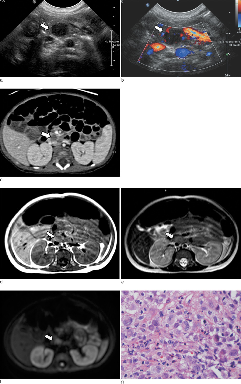
Fig. 1 5-week-old male infant with granulomatous skin lesions. a. Abdominal sonograph showing a round hypoechoic mass (arrow) in the uncinate process of the pancreas. b. Doppler sonograph showing internal vascularity in the pancreatic lesion (arrow). c. Axial image of the abdominal CT scan showing a round lesion with low attenuation and poor enhancement at the uncinate process (arrow) of the pancreas. d-f. Abdominal MRI showing a solid mass (arrow) with low signal intensity on the T1-weighted image (d) and intermediate signal intensity on the T2-weighted image (e). DWI showing the lesion as high signal intensity on the b-value 1000 sec/mm2 image (f), suggestive of diffusion restriction. g. Skin biopsy showing dermal infiltration of histiocytes suggestive of Langerhans cell histiocytosis.
Reference
-
1. Ladisch S JE. Principles and practice of pediatric oncology. 1993. 2nd edition ed. Philadelphia: Lippincott.2. Osband ME, Pochedly C. Histiocytosis-X: an overview. Hematol Oncol Clin North Am. 1987. 1:1–7.3. Favara BE, Feller AC, Pauli M, et al. The WHO Committee On Histiocytic/Reticulum Cell Proliferations. Reclassification Working Group of the Histiocyte Society. Contemporary classification of histiocytic disorders. Med Pediatr Oncol. 1997. 29:157–166.4. Goyal R, Das A, Nijhawan R, Bansal D, Marwaha RK. Langerhans cell histiocytosis infiltration into pancreas and kidney. Pediatr Blood Cancer. 2007. 49:748–750.5. Yu RC, Attra A, Quinn CM, Krausz T, Chu AC. Multisystem Langerhans' cell histiocytosis with pancreatic involvement. Gut. 1993. 34:570–572.6. Muwakkit S, Gharagozloo A, Souid AK, Spirt BA. The sonographic appearance of lesions of the spleen and pancreas in an infant with Langerhans' cell histiocytosis. Pediatr Radiol. 1994. 24:222–223.7. The French Langerhans' Cell Histiocytosis Study Group. A multicentre retrospective survey of Langerhans' cell histiocytosis: 348 cases observed between 1983 and 1993. Arch Dis Child. 1996. 75:17–12.8. Keeling JW, Harries JT. Intestinal malabsorption in infants with histiocytosis X. Arch Dis Child. 1973. 48:350–354.9. Chung EM, Travis MD, Conran RM. Pancreatic tumors in children: radiologic-pathologic correlation. Radiographics. 2006. 26:1211–1238.10. Lahey E. Histiocytosis x--an analysis of prognostic factors. J Pediatr. 1975. 87:184–189.
- Full Text Links
-
- Actions
-
Cited
- CITED
-
- Close
- Share
-
- Similar articles
-
- Langerhans Cell Histiocytosis With Secondary Aneurysmal Bone Cyst in a 9-Year-Old Boy’s Femur: A Case Report
- Isolated Thymic Langerhans Cell Histiocytosis
- Squamous Cell Carcinoma of the Pancreas: A Case Report
- A Case of Gastric Langerhans Cell Histiocytosis Showing Hypertrophic Folds
- Langerhans cell histiocytosis of the mandible: two case reports and literature review
