Isolation and In Vitro Culture of Vascular Endothelial Cells from Mice
- Affiliations
-
- 1Department of Physiology, School of Medicine, Ewha Womans University, Seoul 157-710, Korea. shsuh@ewha.ac.kr
- 2Department of Thoracic & Cardiovascular Surgery and Ewha Womans University Global Top 5 Research Program, School of Medicine, Ewha Womans University, Seoul 157-710, Korea.
- KMID: 2071817
- DOI: http://doi.org/10.4196/kjpp.2015.19.1.35
Abstract
- In cardiovascular disorders, understanding of endothelial cell (EC) function is essential to elucidate the disease mechanism. Although the mouse model has many advantages for in vivo and in vitro research, efficient procedures for the isolation and propagation of primary mouse EC have been problematic. We describe a high yield process for isolation and in vitro culture of primary EC from mouse arteries (aorta, braches of superior mesenteric artery, and cerebral arteries from the circle of Willis). Mouse arteries were carefully dissected without damage under a light microscope, and small pieces of the vessels were transferred on/in a Matrigel matrix enriched with endothelial growth supplement. Primary cells that proliferated in Matrigel were propagated in advanced DMEM with fetal calf serum or platelet-derived serum, EC growth supplement, and heparin. To improve the purity of the cell culture, we applied shearing stress and anti-fibroblast antibody. EC were characterized by a monolayer cobble stone appearance, positive staining with acetylated low density lipoprotein labeled with 1,1'-dioctadecyl-3,3,3',3'-tetramethyl-indocarbocyanine perchlorate, RT-PCR using primers for von-Willebrand factor, and determination of the protein level endothelial nitric oxide synthase. Our simple, efficient method would facilitate in vitro functional investigations of EC from mouse vessels.
Keyword
MeSH Terms
Figure
-

Fig. 1 Pieces of mouse vessels explanted on/in a Matrigel support. Cells started to migrate from the edges of explants and reached confluence. (A) Aortic pieces were placed with the intima side down on Matrigel (left panel) and removed after 5~7 days (middle panel). Higher magnification images of the boxed areas, designated as a and b, are displayed in the right upper and lower sides, respectively. Cells proliferated to form a tube-like structure. (B) Schematic representations of branches of the SMA (left panel) and the CA from the circle of Willis (right panel). Branches of the SMA and the CA from the circle of Willis were explanted in Matrigel, and cells migrated from small segments of the vessels. Images were obtained by phase-contrast microscopy at 50× magnification.

Fig. 2 Labeling of MAEC with DiI-Ac-LDL. (A, B) Active DiI-Ac-LDL uptake was examined by flow cytometry in MAEC at passages 1-5. MAEC at passage 1 without DiI-Ac-LDL were used as a negative control.
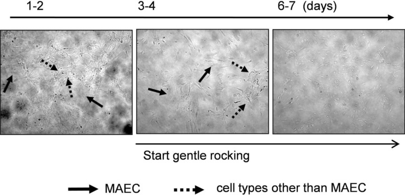
Fig. 3 Elimination of cells in the multilayer. Cells growing in the multilayer were markedly increased after passaging (left panel). When exposed to anti-fibroblast antibody and gentle rocking, cells in the multilayer gradually disappeared. Four or five days after the exposure, only cells growing in a monolayer remained. Lined arrows represent cells in a single layer, and dotted arrows are cells in a multilayer.
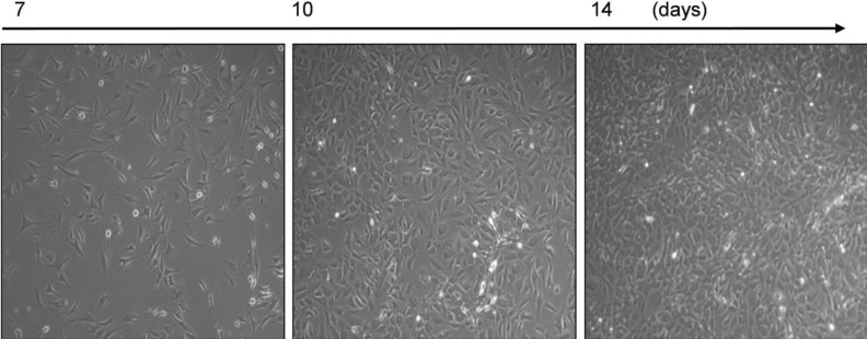
Fig. 4 Purified MAECs by elimination of cells in the multilayer. Cells from aortic explants were exposed to anti-fibroblast antibody and gentle rocking, eliminating cells in the multilayer. A confluent monolayer of MAECs was demonstrated. Note the presence of contact inhibition and cobblestone morphology in all panels. Images were obtained by phase-contrast microscopy at 100× magnification.

Fig. 5 Typical EC characteristics of isolated cells. (A) Cells were stained with DiI-Ac-LDL, and HUVECs were employed as a positive control (left panel). DAPI was used for nucleus staining (center panel), and merged images are displayed (right panel). DiI-Ac-LDL uptake occurred in nearly all of the cells. (B) Flow cytometry analysis was performed in MAECs (passage 4) and HUVEC. DiI-Ac-LDL-labeled ECs showed a dominant fluorescence shift compared to unlabeled negative control cells. (C) Active DiI-Ac-LDL uptake in EC isolated from the CA (CAEC, left panel) and from branches of the SMA (SMAEC, right panel).
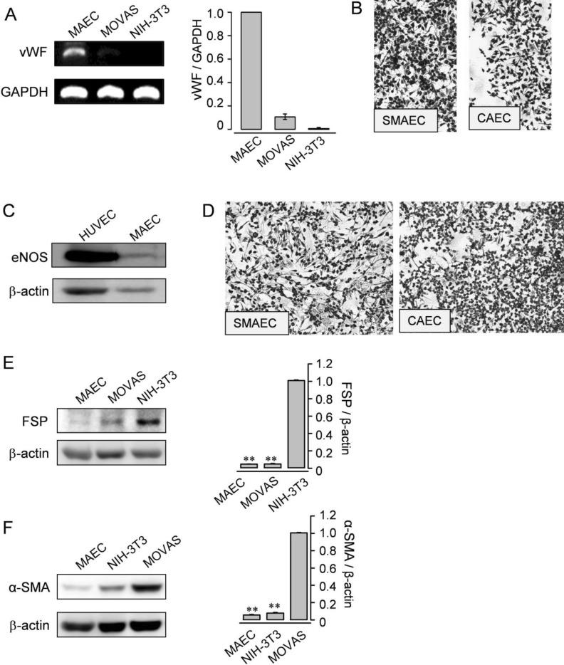
Fig. 6 Expression of typical genes and proteins from ECs. (A) RT-PCR analysis for vWF was performed (passage 5) in MAEC. SMC (MOVAS) and fibroblasts (NIH-3T3) were employed as negative controls. GAPDH was used as an internal standard. (B) Immunocytochemistry of the isolated EC from branches of the SMA or the CA for vWF. (C) Western blotting for eNOS was examined in MAEC (passage 2) and HUVEC. HUVEC were used as a positive control. (D) Immunocytochemistry of the isolated EC from branches of the SMA or the CA for eNOS. (E and F) Western blotting for FSP (E) or α-SMA (F) was examined in MAEC (passage 5), MOVAS, and NIH-3T3. NIH-3T3 or MOVAS was used as a positive control. β-actin was used as an internal standard. (A, E, and F) Results are representative of three independent experiments. The data shown are mean±SEM of three independent experiments.
Reference
-
1. Carmeliet P. Angiogenesis in health and disease. Nat Med. 2003; 9:653–660. PMID: 12778163.
Article2. Choi S, Kim JA, Na HY, Kim JE, Park S, Han KH, Kim YJ, Suh SH. NADPH oxidase 2-derived superoxide downregulates endothelial KCa3.1 in preeclampsia. Free Radic Biol Med. 2013; 57:10–21. PMID: 23261940.
Article3. Choi S, Kim JA, Na HY, Cho SE, Park S, Jung SC, Suh SH. Globotriaosylceramide induces lysosomal degradation of endothelial KCa3.1 in fabry disease. Arterioscler Thromb Vasc Biol. 2014; 34:81–89. PMID: 24158513.4. Park S, Kim JA, Joo KY, Choi S, Choi EN, Shin JA, Han KH, Jung SC, Suh SH. Globotriaosylceramide leads to K(Ca)3.1 channel dysfunction: a new insight into endothelial dysfunction in Fabry disease. Cardiovasc Res. 2011; 89:290–299. PMID: 20971723.
Article5. Ni CW, Kumar S, Ankeny CJ, Jo H. Development of immortalized mouse aortic endothelial cell lines. Vasc Cell. 2014; 6:7. PMID: 24690145.
Article6. Nishiyama T, Mishima K, Ide F, Yamada K, Obara K, Sato A, Hitosugi N, Inoue H, Tsubota K, Saito I. Functional analysis of an established mouse vascular endothelial cell line. J Vasc Res. 2007; 44:138–148. PMID: 17215585.
Article7. Kobayashi M, Inoue K, Warabi E, Minami T, Kodama T. A simple method of isolating mouse aortic endothelial cells. J Atheroscler Thromb. 2005; 12:138–142. PMID: 16020913.
Article8. Magid R, Martinson D, Hwang J, Jo H, Galis ZS. Optimization of isolation and functional characterization of primary murine aortic endothelial cells. Endothelium. 2003; 10:103–109. PMID: 12791518.
Article9. Kevil CG, Bullard DC. In vitro culture and characterization of gene targeted mouse endothelium. Acta Physiol Scand. 2001; 173:151–157. PMID: 11678738.
Article10. Suh SH, Vennekens R, Manolopoulos VG, Freichel M, Schweig U, Prenen J, Flockerzi V, Droogmans G, Nilius B. Characterisation of explanted endothelial cells from mouse aorta: electrophysiology and Ca2+ signalling. Pflugers Arch. 1999; 438:612–620. PMID: 10555557.11. Dong QG, Bernasconi S, Lostaglio S, De Calmanovici RW, Martin-Padura I, Breviario F, Garlanda C, Ramponi S, Mantovani A, Vecchi A. A general strategy for isolation of endothelial cells from murine tissues. Characterization of two endothelial cell lines from the murine lung and subcutaneous sponge implants. Arterioscler Thromb Vasc Biol. 1997; 17:1599–1604. PMID: 9301641.12. Voyta JC, Via DP, Butterfield CE, Zetter BR. Identification and isolation of endothelial cells based on their increased uptake of acetylated-low density lipoprotein. J Cell Biol. 1984; 99:2034–2040. PMID: 6501412.
Article13. Gerritsen ME, Shen CP, McHugh MC, Atkinson WJ, Kiely JM, Milstone DS, Luscinskas FW, Gimbrone MA Jr. Activationdependent isolation and culture of murine pulmonary microvascular endothelium. Microcirculation. 1995; 2:151–163. PMID: 7497167.
Article14. Sahagun G, Moore SA, Fabry Z, Schelper RL, Hart MN. Purification of murine endothelial cell cultures by flow cytometry using fluorescein-labeled griffonia simplicifolia agglutinin. Am J Pathol. 1989; 134:1227–1232. PMID: 2757116.15. Alroy J, Goyal V, Skutelsky E. Lectin histochemistry of mammalian endothelium. Histochemistry. 1987; 86:603–607. PMID: 3610672.
Article16. Gerritsen ME, Shen CP, Atkinson WJ, Padgett RC, Gimbrone MA Jr, Milstone DS. Microvascular endothelial cells from E-selectin-deficient mice form tubes in vitro. Lab Invest. 1996; 75:175–184. PMID: 8765318.17. Barkalow FJ, Goodman MJ, Mayadas TN. Cultured murine cerebral microvascular endothelial cells contain von Willebrand factor-positive Weibel-Palade bodies and support rapid cytokine-induced neutrophil adhesion. Microcirculation. 1996; 3:19–28. PMID: 8846268.18. Barkalow FJ, Goodman MJ, Gerritsen ME, Mayadas TN. Brain endothelium lack one of two pathways of P-selectin-mediated neutrophil adhesion. Blood. 1996; 88:4585–4593. PMID: 8977250.
Article19. Su X, Sorenson CM, Sheibani N. Isolation and characterization of murine retinal endothelial cells. Mol Vis. 2003; 9:171–178. PMID: 12740568.20. Reiss Y, Hoch G, Deutsch U, Engelhardt B. T cell interaction with ICAM-1-deficient endothelium in vitro: essential role for ICAM-1 and ICAM-2 in transendothelial migration of T cells. Eur J Immunol. 1998; 28:3086–3099. PMID: 9808177.
- Full Text Links
-
- Actions
-
Cited
- CITED
-
- Close
- Share
-
- Similar articles
-
- In Vitro Culture of Endothelial Cell and Smooth Muscle Cell for Studying Vascular Diseases
- Effects of Dimethyl Sulfoxide on the Differentiation of Myocardial and Endothelial Cells
- Comparative Evaluation for Potential Differentiation of Endothelial Progenitor Cells and Mesenchymal Stem Cells into Endothelial-Like Cells
- An Effective Isolation of the Vascular Endothelial and Smooth Muscle Cells from the Mouse Aorta
- Culture of the Human Glomerular Endothelial Cells
