Salvage Therapy from Traumatic Ischemic Finger Necrosis via Prostaglandin E1 Assisted Conservative Treatment: A Case Report
- Affiliations
-
- 1Department of Orthopaedic Surgery, Hallym University Dongtan Sacred Heart Hospital, Hwaseong, Korea. jshin2100@gmail.com
- 2Department of Orthopaedics and Traumatology, Cheju Halla General Hospital, Jeju, Korea.
- KMID: 2070351
- DOI: http://doi.org/10.12671/jkfs.2015.28.4.245
Abstract
- Prostaglandin E1 (PGE-1) is a potent vasodilator, which also inhibits platelet aggregation, affects the blood flow viscosity, and fibrinolysis. The compound also excerts anti-inflammatory effects by inhibiting the monocyte and neutrophil function. PGE-1 has been widely administered following microvascular flap surgery, along with perioperative antithrombotic agents such as low molecular weight heparin or aspirin, showing excellent results. We report a case showing successful salvage recovery from post-traumatic ischemic necrosis of the finger via PGE-1 assisted conservative treatment.
Keyword
MeSH Terms
Figure
-
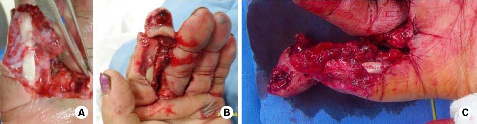
Fig. 1 Initial crushing injury to the index finger. Circulation and sensation were negative at the distal phalangeal part of the injured finger. (A, B) Preoperative image. (C) Intraoperative image.
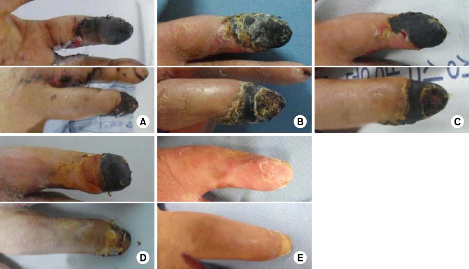
Fig. 2 At postoperative two weeks, the index finger showed gangrenous necrosis distally from the proximal interphalangeal joint level. During the follow-up period with prostaglandin E1 assisted conservative treatment at postoperative 7 weeks, at 8 weeks, and at 11 weeks, the wound healing proceeded, and the necrotic tissue decreased. At postoperative 14 weeks, the Salvage-therapy reconstruction was complete. (A) 2 weeks, (B) 7 weeks, (C) 8 weeks, (D) 11 weeks, and (E) 14 weeks, postoperatively.
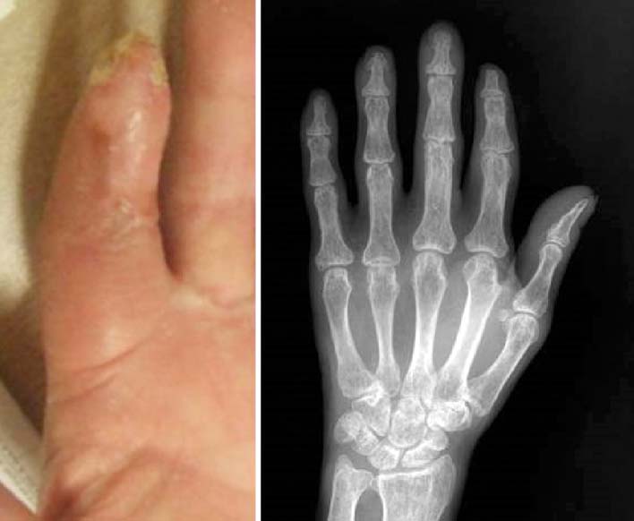
Fig. 3 At postoperative 4 months, the finger was Salvage-reconstructed. Pulp hypotrophy was noted but the partially maintained active range of motion enabled active cupholding (see the main text).
Reference
-
1. Brent B. Replantation of amputated distal phalangeal parts of fingers without vascular anastomoses, using subcutaneous pockets. Plast Reconstr Surg. 1979; 63:1–8.
Article2. Lee JY, Teoh LC, Seah VW. Extending the reach of the heterodigital arterialized flap by cross-finger transfer. Plast Reconstr Surg. 2006; 117:2320–2328.
Article3. Rodríguez Vegas JM, Ruiz Alonso ME, Terán Saavedra PP. PGE-1 in replantation and free tissue transfer: early preliminary experience. Microsurgery. 2007; 27:395–397.
Article4. Matsudaira K, Seichi A, Kunogi J, et al. The efficacy of prostaglandin E1 derivative in patients with lumbar spinal stenosis. Spine (Phila Pa 1976). 2009; 34:115–120.
Article5. Levy JM, Joseph RB, Bodell LS, Nykamp PW, Hessel SJ. Prostaglandin E1 in hand angiography. AJR Am J Roentgenol. 1983; 141:1043–1046.
Article6. Weiner R, Kaley G. Influence of prostaglandin E1 on the terminal vascular bed. Am J Physiol. 1969; 217:563–566.
Article7. Rowlands TE, Gough MJ, Homer-Vanniasinkam S. Do prostaglandins have a salutary role in skeletal muscle ischaemia-reperfusion injury? Eur J Vasc Endovasc Surg. 1999; 18:439–444.
Article8. Campion T, Lynch TG, Kerr JC, Hobson RW 2nd. Effects of prostacyclin injections and infusions on canine femoral hemodynamics. J Vasc Surg. 1986; 3:540–544.
Article9. Iversen VV, Reed RK. PGE1 induced transcapillary transport of 51Cr-EDTA in rat skin measured by microdialysis. Acta Physiol Scand. 2002; 176:269–274.
Article10. Cavadas PC. Supramicrosurgical ear replantation: case report. J Reconstr Microsurg. 2002; 18:393–395.
Article
- Full Text Links
-
- Actions
-
Cited
- CITED
-
- Close
- Share
-
- Similar articles
-
- Effects of Prostaglandin E1 and Supplemental Oxygen on the Wound Healing
- Necrosis of the Penis with Multiple Vessel Atherosclerosis
- Prostaglandin E1 Monotherapy for Impotence
- Intra-arterial infusion of prostaglandin E1 in Buerger's disease: report of 3 cases
- A Case of Intracavernous Needle Breakage during Intracavernous Self-injection of Prostaglandin E1 ( Caverject? )
