Attenuation-Based Automatic Kilovoltage Selection and Sinogram-Affirmed Iterative Reconstruction: Effects on Radiation Exposure and Image Quality of Portal-Phase Liver CT
- Affiliations
-
- 1Department of Radiology, Chonbuk National University Medical School and Hospital, Biomedical Research Institute of Chonbuk National University Hospital, Jeonju 561-712, Korea. pichgo@gmail.com
- KMID: 2069985
- DOI: http://doi.org/10.3348/kjr.2015.16.1.69
Abstract
OBJECTIVE
To compare the radiation dose and image quality between standard-dose CT and a low-dose CT obtained with the combined use of an attenuation-based automatic kilovoltage (kV) selection tool (CARE kV) and sinogram-affirmed iterative reconstruction (SAFIRE) for contrast-enhanced CT examination of the liver.
MATERIALS AND METHODS
We retrospectively reviewed 67 patients with chronic liver disease in whom both, standard-dose CT with 64-slice multidetector-row CT (MDCT) (protocol A), and low-dose CT with 128-slice MDCT using CARE kV and SAFIRE (protocol B) were performed. Images from protocol B during the portal phase were reconstructed using either filtered back projection or SAFIRE with 5 different iterative reconstruction (IR) strengths. We performed qualitative and quantitative analyses to select the appropriate IR strength. Reconstructed images were then qualitatively and quantitatively compared with protocol A images.
RESULTS
Qualitative and quantitative analysis of protocol B demonstrated that SAFIRE level 2 (S2) was most appropriate in our study. Qualitative and quantitative analysis comparing S2 images from protocol B with images from protocol A, showed overall good diagnostic confidence of S2 images despite a significant radiation dose reduction (47% dose reduction, p < 0.001).
CONCLUSION
Combined use of CARE kV and SAFIRE allowed significant reduction in radiation exposure while maintaining image quality in contrast-enhanced liver CT.
Keyword
MeSH Terms
Figure
-

Fig. 1 Transverse contrast-enhanced liver CT images of 47-year-old female (body mass index, 22.4 kg/m2) with chronic hepatitis B. All images were obtained with tube voltage of 100 kV and 110 effective mAs (protocol B). Image noise decreased as SAFIRE level increased. However, as level increased, pixelated image appearance also increased.
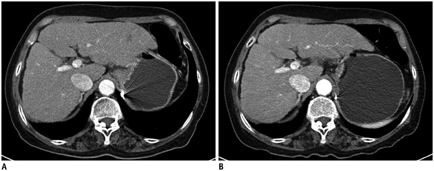
Fig. 2 Transverse contrast-enhanced liver CT images of 67-year-old female (body mass index, 26.7 kg/m2) with liver cirrhosis. Previous CT (A) was scanned at 120 kV (136 eff. mAs) with volume CT dose index (CTDIvol) of 10.4 mGy and follow-up CT (B) was performed at 100 kV (132 eff. mAs) with CTDIvol of 5.2 mGy. Both readers selected (B) as preferred image.
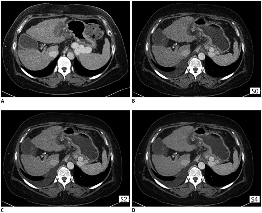
Fig. 3 Transverse contrast-enhanced liver CT images of 52-year-old female (body mass index, 33.2 kg/m2) with liver cirrhosis and history of radiofrequency ablation due to hepatocellular carcinoma. Previous CT (A) was scanned at 120 kV (249 eff. mAs) with volume CT dose index (CTDIvol) of 16.48 mGy and follow-up CT (B-D) was scanned at 100 kV (244 eff. mAs) with CTDIvol of 9.6 mGy. Although S4 (D) and S5 (not shown) images are almost free of image noise, overall diagnostic confidence was rated average (score 3) by both readers due to pixelated and artificial appearance. For comparison of protocol A and B, both readers selected 2 images (A, C) with same preference.
Reference
-
1. Lee KH, Lee JM, Moon SK, Baek JH, Park JH, Flohr TG, et al. Attenuation-based automatic tube voltage selection and tube current modulation for dose reduction at contrast-enhanced liver CT. Radiology. 2012; 265:437–447.2. Brenner DJ, Hall EJ. Computed tomography--an increasing source of radiation exposure. N Engl J Med. 2007; 357:2277–2284.3. Fazel R, Krumholz HM, Wang Y, Ross JS, Chen J, Ting HH, et al. Exposure to low-dose ionizing radiation from medical imaging procedures. N Engl J Med. 2009; 361:849–857.4. Mulkens TH, Bellinck P, Baeyaert M, Ghysen D, Van Dijck X, Mussen E, et al. Use of an automatic exposure control mechanism for dose optimization in multi-detector row CT examinations: clinical evaluation. Radiology. 2005; 237:213–223.5. McCollough CH, Bruesewitz MR, Kofler JM Jr. CT dose reduction and dose management tools: overview of available options. Radiographics. 2006; 26:503–512.6. Goo HW. CT radiation dose optimization and estimation: an update for radiologists. Korean J Radiol. 2012; 13:1–11.7. McCollough CH, Primak AN, Braun N, Kofler J, Yu L, Christner J. Strategies for reducing radiation dose in CT. Radiol Clin North Am. 2009; 47:27–40.8. Yu L, Bruesewitz MR, Thomas KB, Fletcher JG, Kofler JM, McCollough CH. Optimal tube potential for radiation dose reduction in pediatric CT: principles, clinical implementations, and pitfalls. Radiographics. 2011; 31:835–848.9. Marin D, Nelson RC, Schindera ST, Richard S, Youngblood RS, Yoshizumi TT, et al. Low-tube-voltage, high-tube-current multidetector abdominal CT: improved image quality and decreased radiation dose with adaptive statistical iterative reconstruction algorithm--initial clinical experience. Radiology. 2010; 254:145–153.10. Yu L, Li H, Fletcher JG, McCollough CH. Automatic selection of tube potential for radiation dose reduction in CT: a general strategy. Med Phys. 2010; 37:234–243.11. Siegel MJ, Schmidt B, Bradley D, Suess C, Hildebolt C. Radiation dose and image quality in pediatric CT: effect of technical factors and phantom size and shape. Radiology. 2004; 233:515–522.12. Schindera ST, Winklehner A, Alkadhi H, Goetti R, Fischer M, Gnannt R, et al. Effect of automatic tube voltage selection on image quality and radiation dose in abdominal CT angiography of various body sizes: a phantom study. Clin Radiol. 2013; 68:e79–e86.13. Sigal-Cinqualbre AB, Hennequin R, Abada HT, Chen X, Paul JF. Low-kilovoltage multi-detector row chest CT in adults: feasibility and effect on image quality and iodine dose. Radiology. 2004; 231:169–174.14. Schabel C, Fenchel M, Schmidt B, Flohr TG, Wuerslin C, Thomas C, et al. Clinical evaluation and potential radiation dose reduction of the novel sinogram-affirmed iterative reconstruction technique (SAFIRE) in abdominal computed tomography angiography. Acad Radiol. 2013; 20:165–172.15. Shin HJ, Chung YE, Lee YH, Choi JY, Park MS, Kim MJ, et al. Radiation dose reduction via sinogram affirmed iterative reconstruction and automatic tube voltage modulation (CARE kV) in abdominal CT. Korean J Radiol. 2013; 14:886–893.16. Eller A, May MS, Scharf M, Schmid A, Kuefner M, Uder M, et al. Attenuation-based automatic kilovolt selection in abdominal computed tomography: effects on radiation exposure and image quality. Invest Radiol. 2012; 47:559–565.17. Winklehner A, Goetti R, Baumueller S, Karlo C, Schmidt B, Raupach R, et al. Automated attenuation-based tube potential selection for thoracoabdominal computed tomography angiography: improved dose effectiveness. Invest Radiol. 2011; 46:767–773.18. Gnannt R, Winklehner A, Eberli D, Knuth A, Frauenfelder T, Alkadhi H. Automated tube potential selection for standard chest and abdominal CT in follow-up patients with testicular cancer: comparison with fixed tube potential. Eur Radiol. 2012; 22:1937–1945.19. Kalra MK, Woisetschläger M, Dahlström N, Singh S, Lindblom M, Choy G, et al. Radiation dose reduction with Sinogram Affirmed Iterative Reconstruction technique for abdominal computed tomography. J Comput Assist Tomogr. 2012; 36:339–346.20. Baker ME, Dong F, Primak A, Obuchowski NA, Einstein D, Gandhi N, et al. Contrast-to-noise ratio and low-contrast object resolution on full- and low-dose MDCT: SAFIRE versus filtered back projection in a low-contrast object phantom and in the liver. AJR Am J Roentgenol. 2012; 199:8–18.21. Pontana F, Pagniez J, Duhamel A, Flohr T, Faivre JB, Murphy C, et al. Reduced-dose low-voltage chest CT angiography with Sinogram-affirmed iterative reconstruction versus standard-dose filtered back projection. Radiology. 2013; 267:609–618.22. Singh S, Kalra MK, Moore MA, Shailam R, Liu B, Toth TL, et al. Dose reduction and compliance with pediatric CT protocols adapted to patient size, clinical indication, and number of prior studies. Radiology. 2009; 252:200–208.23. Awai K, Takada K, Onishi H, Hori S. Aortic and hepatic enhancement and tumor-to-liver contrast: analysis of the effect of different concentrations of contrast material at multi-detector row helical CT. Radiology. 2002; 224:757–763.24. Spielmann AL, Nelson RC, Lowry CR, Johnson GA, Sundaramoothy G, Sheafor DH, et al. Liver: single breath-hold dynamic subtraction CT with multi-detector row helical technology feasibility study. Radiology. 2002; 222:278–283.25. CT Dosimetry Diagnostic Imaging Council CT Committee. AAPM REPORT NO. 96. The Measurement, Reporting, and Management of Radiation Dose in CT. USA: American Association of Physicists in Medicine;2008.26. Conover WJ. The Friedman test. In : Conover WJ, editor. Practical Nonparametric Statistics. New York: J. Wiley;1999. p. 369–373.27. Schindera ST, Nelson RC, Yoshizumi T, Toncheva G, Nguyen G, DeLong DM, et al. Effect of automatic tube current modulation on radiation dose and image quality for low tube voltage multidetector row CT angiography: phantom study. Acad Radiol. 2009; 16:997–1002.28. Hur S, Lee JM, Kim SJ, Park JH, Han JK, Choi BI. 80-kVp CT using Iterative Reconstruction in Image Space algorithm for the detection of hypervascular hepatocellular carcinoma: phantom and initial clinical experience. Korean J Radiol. 2012; 13:152–164.29. Namimoto T, Oda S, Utsunomiya D, Shimonobo T, Morita S, Nakaura T, et al. Improvement of image quality at low-radiation dose and low-contrast material dose abdominal CT in patients with cirrhosis: intraindividual comparison of low tube voltage with iterative reconstruction algorithm and standard tube voltage. J Comput Assist Tomogr. 2012; 36:495–501.30. Karpitschka M, Augart D, Becker HC, Reiser M, Graser A. Dose reduction in oncological staging multidetector CT: effect of iterative reconstruction. Br J Radiol. 2013; 86:20120224.
- Full Text Links
-
- Actions
-
Cited
- CITED
-
- Close
- Share
-
- Similar articles
-
- Combined Use of Automatic Tube Voltage Selection and Current Modulation with Iterative Reconstruction for CT Evaluation of Small Hypervascular Hepatocellular Carcinomas: Effect on Lesion Conspicuity and Image Quality
- Quantitative Evaluation of Cardiothoracic CT Image Sharpness with an Integrated Circuit Detector: Comparison between Statistical and Model-Based Iterative Reconstruction Algorithms
- Radiation Dose Reduction via Sinogram Affirmed Iterative Reconstruction and Automatic Tube Voltage Modulation (CARE kV) in Abdominal CT
- Effects of Iterative Reconstruction Algorithm, Automatic Exposure Control on Image Quality, and Radiation Dose: Phantom Experiments with Coronary CT Angiography Protocols
- Attenuation-Based Automatic Tube Potential Selection in Cerebral Computed Tomography Angiography: Effects on Radiation Exposure and Image Quality
