Multidetector Computed Tomography for the Evaluation of Coronary Artery Disease; The Diagnostic Accuracy in Calcified Coronary Arteries, Comparing with IVUS Imaging
- Affiliations
-
- 1Division of Cardiology, Department of Internal Medicine, NHIS Ilsan Hospital, Goyang, Korea.
- 2Division of Cardiology, Department of Internal Medicine, Heart Center, Gangnam Severance Hospital, Yonsei University College of Medicine, Seoul, Korea. cardiobk@yuhs.ac
- 3Department of Radiology, Gangnam Severance Hospital, Yonsei University College of Medicine, Seoul, Korea.
- 4Division of Cardiology, Department of Internal Medicine, Yongin Severance Hospital, Yonsei University College of Medicine, Yongin, Korea.
- KMID: 2068668
- DOI: http://doi.org/10.3349/ymj.2014.55.3.599
Abstract
- PURPOSE
Contrast enhanced multidetector computed tomography (MDCT) has been used as an alternative to coronary angiography for the assessment of coronary artery disease in the patient of the intermediate risk group. However, coronary calcium is a known limiting factor for MDCT evaluation. We investigated the diagnostic accuracy of 64-channel MDCT with each coronary artery calcium score (CACS) by compared with intravascular ultrasound (IVUS) imaging.
MATERIALS AND METHODS
A total of 54 symptomatic patients with intermediate-risk (10 females, mean age 59.9+/-6.9 years, Framingham point scores 9-20) with 162 sites who had a culprit lesion on 64-channel MDCT before performing coronary angiography with IVUS were enrolled. Patients were divided into 4 subgroups depending on CACS: 0, 1-99, 100-399, and >400. Lesion length, external elastic membrane (EEM) cross sectional area (CSA), minimal luminal area, and plaque area were measured and compared between IVUS and MDCT.
RESULTS
The correlation coefficients for the measurements of the EEM CSA, lumen CSA, and plaque area were r=0.514, r=0.837, and r=0.578, respectively. Furthermore, there were close correlation of plaque area between four subgroups of CACS (r=0.671, r=0.623, r=0.562, r=0.571, respectively).
CONCLUSION
Despite the increase in CACS, the geometric analysis of coronary arteries using with 64-channel MDCT was comparable with IVUS in symptomatic patient of the intermediate risk group.
Keyword
MeSH Terms
Figure
-
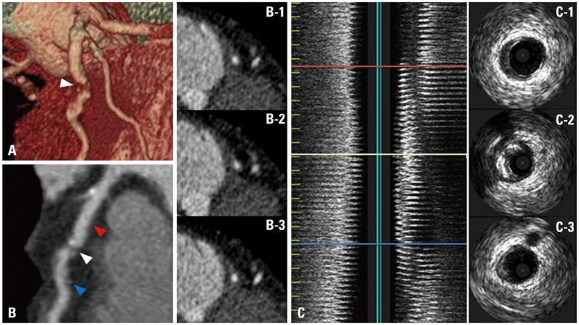
Fig. 1 Coronary plaques in the mid-left anterior descending artery of a patient presenting with stable angina on MDCT (A and B) and IVUS (C). MDCT, multidetector computed tomography; IVUS, intravascular ultrasound.
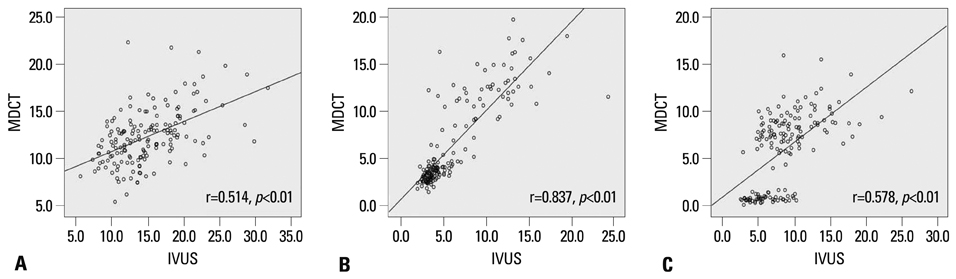
Fig. 2 Linear correlations between IVUS and MDCT measurements. There are significant correlations for EEM CSA (A), lumen CSA (B), and plaque area (C) between them. MDCT, multidetector computed tomography; IVUS, intravascular ultrasound; EEM, external elastic membrane; CSA, cross-sectional area.
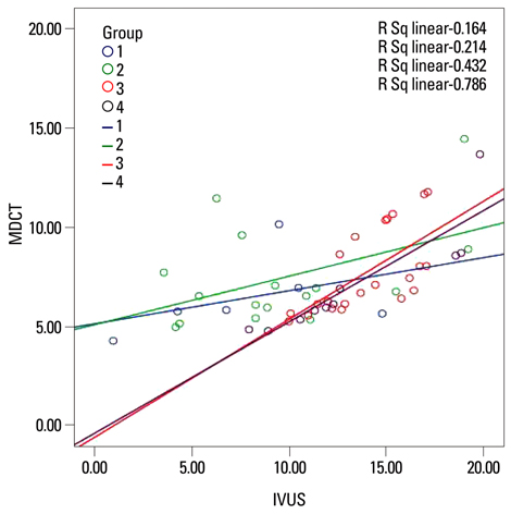
Fig. 3 Linear correlations of plaque area between IVUS and MDCT measurements on each coronary artery calcium group. IVUS, intravascular ultrasound; MDCT, multidetector computed tomography.
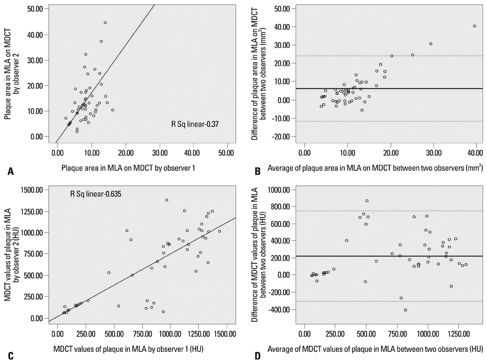
Fig. 4 Inter-observer variability between two observers of plaque area in MLA on MDCT (A), Bland and Altman plot (B) and MDCT values of plaque in MLA (C), Bland and Altman plot (D). MLA, minimal luminal area; HU, Hounsfield unit; MDCT, multidetector computed tomography.
Cited by 1 articles
-
Assessment of Coronary Artery Calcium Scoring for Statin Treatment Strategy according to ACC/AHA Guidelines in Asymptomatic Korean Adults
Donghee Han, Bríain Ó Hartaigh, Ji Hyun Lee, Asim Rizvi, Hyo Eun Park, Su-Yeon Choi, Jidong Sung, Hyuk-Jae Chang
Yonsei Med J. 2017;58(1):82-89. doi: 10.3349/ymj.2017.58.1.82.
Reference
-
1. Falk E, Shah PK, Fuster V. Coronary plaque disruption. Circulation. 1995; 92:657–671.
Article2. Fayad ZA, Fuster V. Clinical imaging of the high-risk or vulnerable atherosclerotic plaque. Circ Res. 2001; 89:305–316.
Article3. Muller JE, Abela GS, Nesto RW, Tofler GH. Triggers, acute risk factors and vulnerable plaques: the lexicon of a new frontier. J Am Coll Cardiol. 1994; 23:809–813.
Article4. Gibbons RJ, Chatterjee K, Daley J, Douglas JS, Fihn SD, Gardin JM, et al. ACC/AHA/ACP-ASIM guidelines for the management of patients with chronic stable angina: a report of the American College of Cardiology/American Heart Association Task Force on Practice Guidelines (Committee on Management of Patients With Chronic Stable Angina). J Am Coll Cardiol. 1999; 33:2092–2197.5. Inoue F, Sato Y, Matsumoto N, Tani S, Uchiyama T. Evaluation of plaque texture by means of multislice computed tomography in patients with acute coronary syndrome and stable angina. Circ J. 2004; 68:840–844.
Article6. Achenbach S, Moselewski F, Ropers D, Ferencik M, Hoffmann U, MacNeill B, et al. Detection of calcified and noncalcified coronary atherosclerotic plaque by contrast-enhanced, submillimeter multidetector spiral computed tomography: a segment-based comparison with intravascular ultrasound. Circulation. 2004; 109:14–17.
Article7. Leber AW, Knez A, von Ziegler F, Becker A, Nikolaou K, Paul S, et al. Quantification of obstructive and nonobstructive coronary lesions by 64-slice computed tomography: a comparative study with quantitative coronary angiography and intravascular ultrasound. J Am Coll Cardiol. 2005; 46:147–154.
Article8. Vanhoenacker PK, Heijenbrok-Kal MH, Van Heste R, Decramer I, Van Hoe LR, Wijns W, et al. Diagnostic performance of multidetector CT angiography for assessment of coronary artery disease: meta-analysis. Radiology. 2007; 244:419–428.
Article9. Budoff MJ, Dowe D, Jollis JG, Gitter M, Sutherland J, Halamert E, et al. Diagnostic performance of 64-multidetector row coronary computed tomographic angiography for evaluation of coronary artery stenosis in individuals without known coronary artery disease: results from the prospective multicenter ACCURACY (Assessment by Coronary Computed Tomographic Angiography of Individuals Undergoing Invasive Coronary Angiography) trial. J Am Coll Cardiol. 2008; 52:1724–1732.
Article10. Naghavi M, Falk E, Hecht HS, Jamieson MJ, Kaul S, Berman D, et al. From vulnerable plaque to vulnerable patient--Part III: Executive summary of the Screening for Heart Attack Prevention and Education (SHAPE) Task Force report. Am J Cardiol. 2006; 98:2H–15H.
Article11. Hoffmann U, Moselewski F, Cury RC, Ferencik M, Jang IK, Diaz LJ, et al. Predictive value of 16-slice multidetector spiral computed tomography to detect significant obstructive coronary artery disease in patients at high risk for coronary artery disease: patient-versus segment-based analysis. Circulation. 2004; 110:2638–2643.
Article12. Ong TK, Chin SP, Liew CK, Chan WL, Seyfarth MT, Liew HB, et al. Accuracy of 64-row multidetector computed tomography in detecting coronary artery disease in 134 symptomatic patients: influence of calcification. Am Heart J. 2006; 151:1323.
Article13. Anderson KM, Odell PM, Wilson PW, Kannel WB. Cardiovascular disease risk profiles. Am Heart J. 1991; 121(1 Pt 2):293–298.
Article14. Wilson PW, Castelli WP, Kannel WB. Coronary risk prediction in adults (the Framingham Heart Study). Am J Cardiol. 1987; 59:91G–94G.
Article15. Mintz GS, Nissen SE, Anderson WD, Bailey SR, Erbel R, Fitzgerald PJ, et al. American College of Cardiology Clinical Expert Consensus Document on Standards for Acquisition, Measurement and Reporting of Intravascular Ultrasound Studies (IVUS). A report of the American College of Cardiology Task Force on Clinical Expert Consensus Documents. J Am Coll Cardiol. 2001; 37:1478–1492.
Article16. Carlier SG, Mintz GS, Stone GW. Imaging of atherosclerotic plaque using radiofrequency ultrasound signal processing. J Nucl Cardiol. 2006; 13:831–840.
Article17. Jakob M, Spasojevic D, Krogmann ON, Wiher H, Hug R, Hess OM. Tortuosity of coronary arteries in chronic pressure and volume overload. Cathet Cardiovasc Diagn. 1996; 38:25–31.
Article18. Moselewski F, Ropers D, Pohle K, Hoffmann U, Ferencik M, Chan RC, et al. Comparison of measurement of cross-sectional coronary atherosclerotic plaque and vessel areas by 16-slice multidetector computed tomography versus intravascular ultrasound. Am J Cardiol. 2004; 94:1294–1297.
Article19. Leber AW, Becker A, Knez A, von Ziegler F, Sirol M, Nikolaou K, et al. Accuracy of 64-slice computed tomography to classify and quantify plaque volumes in the proximal coronary system: a comparative study using intravascular ultrasound. J Am Coll Cardiol. 2006; 47:672–677.
Article20. Motoyama S, Sarai M, Harigaya H, Anno H, Inoue K, Hara T, et al. Computed tomographic angiography characteristics of atherosclerotic plaques subsequently resulting in acute coronary syndrome. J Am Coll Cardiol. 2009; 54:49–57.
Article21. Hein PA, Romano VC, Lembcke A, May J, Rogalla P. Initial experience with a chest pain protocol using 320-slice volume MDCT. Eur Radiol. 2009; 19:1148–1155.
Article
- Full Text Links
-
- Actions
-
Cited
- CITED
-
- Close
- Share
-
- Similar articles
-
- Assessment of Non-Calcified Coronary Plaques Using 64-Slice Computed Tomography: Comparison With Intravascular Ultrasound
- Assessment of Coronary Artery Bypass Graft Patency Using Multidetector Computed Tomography
- Coronary Angiography with Multidetector row Computed Tomography: Part II - Clinical Aspects
- Coronary CT Angiography
- A Single Coronary Artery: Right Coronary Artery Originating From the Distal Left Circumflex Artery
