Assessment of Non-Calcified Coronary Plaques Using 64-Slice Computed Tomography: Comparison With Intravascular Ultrasound
- Affiliations
-
- 1Department of Cardiology, College of Medicine, Catholic University of Daegu, Daegu, Korea. kks7379@cu.ac.kr
- KMID: 1825965
- DOI: http://doi.org/10.4070/kcj.2009.39.3.95
Abstract
-
BACKGROUND AND OBJECTIVES: Non-invasive detection and characterization of plaque composition may constitute an important step in risk stratification and monitoring of the progression of coronary atherosclerosis. Multislice computed tomography (MSCT) allows for accurate, non-invasive detection and characterization of atherosclerotic plaques, as well as determination of coronary artery stenosis. The aim of this study was to determine the usefulness of MSCT for characterizing non-calcified coronary plaques previously classified by intravascular ultrasound (IVUS).
SUBJECTS AND METHODS
Seventy-one plaques were evaluated in 42 patients undergoing MSCT and IVUS. Coronary plaques were classified as hypoechoic or hyperechoic based on IVUS echogenicity. On MSCT, CT attenuation was measured using circular regions of interest (ROI) and represented as Hounsfield units (HU).
RESULTS
MSCT attenuation in hypoechoic plaques was significantly lower than it was in hyperechoic plaques (52.9+/-24.6 HU vs. 98.6+/-34.9 HU, respectively, p<0.001). When comparing CT attenuation between hypoechoic and hyperechoic plaques, 60.2 HU was the cut-off value for differentiating between the two, with a 90.7% sensitivity and a 78.6% specificity.
CONCLUSION
MSCT might be a useful tool for non-invasively evaluating the characteristics of coronary artery plaques.
MeSH Terms
Figure
-

Fig. 1 Measurement of CT attenuation (A) in the plaque corresponding to IVUS (B) (arrow). The CT attenuation data was based on the mean value in different three sites. IVUS: intravascular ultrasound, CT: computed tomography
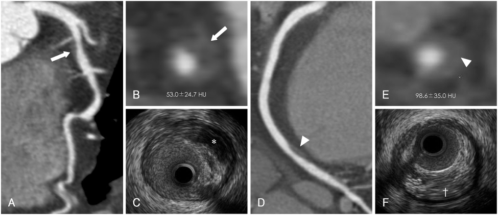
Fig. 2 Visualization of non-calcified plaques by MSCT and IVUS. The CT attenuation (arrow of A and B) of hypoechoic plaques on IVUS (*, C) was lower than the CT attenuation (arrow head of D and E) of hyperechoic plaques (†, F). MSCT: multislice computed tomography, IVUS: intravascular ultrasound, HU: Hounsfield units.
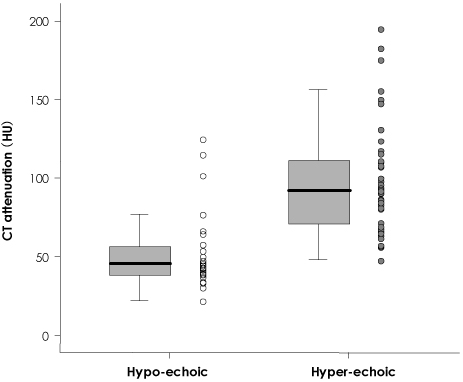
Fig. 3 Comparison of CT attenuation according to plaque echogenicity on IVUS. IVUS: intravascular ultrasound, HU: Hounsfield units.
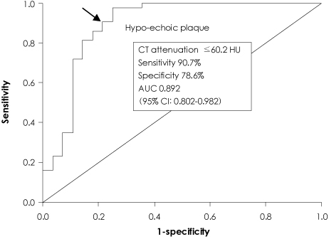
Fig. 4 A receiver operating characteristic curve of CT attenuation for predicting hypo-echoic plaques on IVUS. AUC: area under the curve, IVUS: intravascular ultrasound, CI: confidence interval.
Reference
-
1. Motoyama S, Kondo T, Anno H, et al. Atherosclerotic plaque characterization by 0.5 mm-slice multislice computed tomographic imaging. Circ J. 2007. 71:363–366.2. Kunimasa T, Sato Y, Sugi K, Moroi M. Evaluation by multislice computed tomography of atherosclerotic coronary artery plaques in non-culprit, remote coronary arteries of patients with acute coronary syndorme. Circ J. 2005. 69:1346–1351.3. Kim KS. Imaging markers of subclinical atherosclerosis. Korean Circ J. 2007. 37:1–8.4. Schroder S, Kopp AF, Baumbach A, et al. Noninvasive detection and evaluation of atherosclerotic coronary plaques with multislice computed tomography. J Am Coll Cardiol. 2001. 37:1430–1435.5. Leber AW, Knez A, Becker A, et al. Accuracy of multidetector spiral computed tomography in identifying and differentiating the composition of coronary atherosclerotic plaques: a comparative study with intracoronary ultrasound. J Am Coll Cardiol. 2004. 43:1241–1247.6. Estes JM, Quist WC, Lo Gerfo FW, Costello P. Noninvasive characterization of plaque morphology using helical computed tomography. J Cardiovasc Surg (Torino). 1998. 39:527–534.7. Pohle K, Achenbach S, Macneill B, et al. Characterization of non-calcified coronary atherosclerotic plaque by multi-detector row CT: comparison to IVUS. Atherosclerosis. 2007. 190:174–180.8. Davies MJ, Thomas AC. Plaque fissuring: the cause of acute myocardial infarction, sudden ischaemic death, and crescendo angina. Br Heart J. 1985. 53:363–373.9. Toussaint JF, LaMuraglia GM, Southern JF, Fuster V, Kantor HL. Magnetic reasonance images of lipid, fibrous, calcified, hemorrhagic and thrombotic components of human atherosclerosis in vivo. Circulation. 1996. 94:932–938.10. Becker CR, Nikolaou K, Muders M, et al. Ex vivo coronary atherosclerotic plaque characterization with multi-detector-row CT. Eur Radiol. 2003. 13:2094–2098.11. Yamagishi M, Terashima M, Awano K, et al. Morphology of vulnerable coronary plaque: insights from follow-up of patients examined by intravascular ultrasound before an acute coronary syndrome. J Am Coll Cardiol. 2000. 35:106–111.12. Funabashi N, Misumi K, Ohnishi H, Asano M, Komuro I. Characterization and morphology of atherosclerotic plaque of coronary arteries: utility of electron-beam tomography to detect non-calcified plaque: a comparison with conventional coronary angiography and intravascular ultrasound. Int J Cardiol. 2007. 115:108–113.13. Rasouli ML, Shavelle DM, French WJ, Mckay CR, Budoff MJ. Assessment of coronary plaque morphology by contrast-enhanced computed tomographic angiography: comparison with intravascular ultrasound. Coron Artery Dis. 2006. 17:359–364.14. Choe YH. Noninvasive imaging of atherosclerotic plaques using MRI and CT. Korean Circ J. 2005. 35:1–14.15. Carrascosa PM, Capunay CM, Merletti PG, Carrascosa J, Garcia JG. Characterization of coronary atherosclerotic plaques by multidetector computed tomography. Am J Cardiol. 2006. 97:598–602.16. Schroeder S, Kiettner A, Leitritz M, et al. Reliability of differentiating human coronary plaque morphology using contrast-enhanced multislice spiral computed tomography. J Comput Assist Tomogr. 2004. 28:449–454.17. Mollet NR, Cademartiri F, Nieman K, et al. Noninvasive assessment of coronary plaque burden using multislice computed tomography. Am J Cardiol. 2005. 95:1165–1169.18. Iriat X, Brunot S, Coste P, et al. Early characterization of atherosclerotic coronary plaques with multidetector computed tomography in patients with acute coronary syndrome: a comparative study with intravascular ultrasound. Eur Radiol. 2007. 17:2581–2588.
- Full Text Links
-
- Actions
-
Cited
- CITED
-
- Close
- Share
-
- Similar articles
-
- Noninvasive Detection of Coronary Atherosclerotic Plaques and Assessment of Stenosis Degree at Multidetector CT Coronary Angiography
- Serial Changes of Coronary Atherosclerotic Plaque: Assessment with 64-Slice Multi-Detector Computed Tomography
- A Case of Coronary Pseudostenosis, Diagnosed by Intravascular Ultrasound
- Nonalcoholic Fatty Liver Disease Is Associated with the Presence and Morphology of Subclinical Coronary Atherosclerosis
- Coronary CT Angiography with Knowledge-Based Iterative Model Reconstruction for Assessing Coronary Arteries and Non-Calcified Predominant Plaques
