Morphometrical Study of Uncinate Processes and Vertebral Body of Cervical Spine
- Affiliations
-
- 1Department of Neurosurgery, 21st Century Hospital, Seoul, Korea.
- 2Department of Neurosurgery, Ewha Womans University School of Medicine, Mokdong Hospital, Seoul, Korea. sjkimmd@unitel.co.kr
- 3Department of Anatomy, Yonsei University School of Medicine, Seoul, Korea.
- KMID: 2066921
- DOI: http://doi.org/10.3340/jkns.2012.51.5.247
Abstract
OBJECTIVE
The anatomical knowledge is the most important and has a direct link with success of operation in cervical spine surgery. The authors measured various cervical parameters in cadaveric dry bones and compared with previous reported results.
METHODS
We made 255 dry bones age from 19 to 72 years (mean, 42.3 years) that were obtained from 51 subjects in 100 subjects who donated their bodies. All measurements from C3-C7 levels were made using digital vernier calipers, standard goniometer, and self-made fix tool for two different cervical axes (canal and disc setting). We classified into 4 groups (uncinate process, vertebral body, lamina, and pedicle) and measured independently by two neurosurgeons for 28 parameters.
RESULTS
We analyzed 23970 measurements by mean value and standard deviations. In comparing with previous literatures, there are some different results. The mean values for uncinate process (UP) width ranged from 5.5 mm at C4 and 5 to 6.3 mm at C3 and C7 in men. Also, in women, the mean values for UP width ranged from 5.5 mm at C5 to 6.3 mm at C7. C7 was widest and C5 was most narrow than other levels. The antero-posterior length of UP tended to increase gradually from C3 to C6. The tip way, tip distance, and base distance of UP also showed increasing pattern from C3 to C7.
CONCLUSION
These measurements can provide the spinal surgeons with a starting point to address bony architectures surrounding targeted soft tissues for safeguard against unintended damages during cervical operation.
Keyword
Figure
-
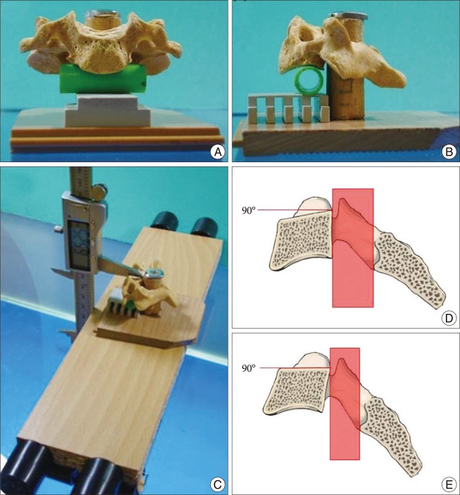
Fig. 1 The self-made fix tools which authors made and used. Anterior (A), lateral (B), and oblique (C) aspect applied with vernier calipers. Schematic sagittal figures of two different axes for cervical vertebra. Canal setting (D) is the state when imaginary longitudinal line made by posterior side of vertebral body is perpendicular to the ground. Disc setting (E) is the state when imaginary horizontal line made by superior side of vertebral body (endplate) is parallel to the ground.
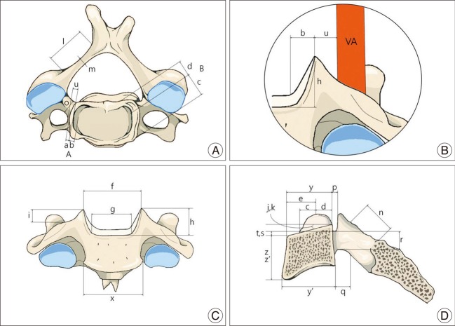
Fig. 2 Schematic figures of parameters of cervical vertebra. Superior (A), enlarged figure of anterior with imaginary line of vertebral artery (B), anterior (C), and sagittal cross section of lateral aspect (D). Illustration (A) from The Textbook of Spine, Korea, 2004, p.37, with permission.
Reference
-
1. Clausen JD, Goel VK, Traynelis VC, Scifert J. Uncinate processes and Luschka joints influence the biomechanics of the cervical spine : quantification using a finite element model of the C5-C6 segment. J Orthop Res. 1997; 15:342–347. PMID: 9246079.
Article2. Ebraheim NA, Lu J, Biyani A, Brown JA, Yeasting RA. Anatomic considerations for uncovertebral involvement in cervical spondylosis. Clin Orthop Relat Res. 1997; 200–206.
Article3. Ebraheim NA, Lu J, Brown JA, Biyani A, Yeasting RA. Vulnerability of vertebral artery in anterolateral decompression for cervical spondylosis. Clin Orthop Relat Res. 1996; 146–151. PMID: 8542690.
Article4. Ebraheim NA, Xu R, Bhatti RA, Yeasting RA. The projection of the cervical disc and uncinate process on the posterior aspect of the cervical spine. Surg Neurol. 1999; 51:363–367. PMID: 10199287.
Article5. Ebraheim NA, Xu R, Lin D, Haman S, Yeasting RA. Quantitative anatomy of the transverse foramen and pedicle of the axis. J Spinal Disord. 1998; 11:521–525. PMID: 9884298.
Article6. Frykholm R. Lower cervical vertebrae and intervertebral discs; surgical anatomy and pathology. Acta Chir Scand. 1951; 101:345–359. PMID: 14868335.7. Lu J, Ebraheim NA, Yang H, Skie M, Yeasting RA. Cervical uncinate process : an anatomic study for anterior decompression of the cervical spine. Surg Radiol Anat. 1998; 20:249–252. PMID: 9787390.
Article8. Marchiori DM, Henderson CN. A cross-sectional study correlating cervical radiographic degenerative findings to pain and disability. Spine (Phila Pa 1976). 1996; 21:2747–2751. PMID: 8979320.
Article9. Milne N. The role of zygapophysial joint orientation and uncinate processes in controlling motion in the cervical spine. J Anat. 1991; 178:189–201. PMID: 1810926.10. Pait TG, Killefer JA, Arnautovic KI. Surgical anatomy of the anterior cervical spine : the disc space, vertebral artery, and associated bony structures. Neurosurgery. 1996; 39:769–776. PMID: 8880772.
Article11. Panjabi MM, Duranceau J, Goel V, Oxland T, Takata K. Cervical human vertebrae. Quantitative three-dimensional anatomy of the middle and lower regions. Spine (Phila Pa 1976). 1991; 16:861–869. PMID: 1948369.
Article12. Payne EE, Spillane JD. The cervical spine; an anatomico-pathological study of 70 specimens (using a special technique) with particular reference to the problem of cervical spondylosis. Brain. 1957; 80:571–596. PMID: 13499761.
Article13. Saringer W, Nöbauer I, Reddy M, Tschabitscher M, Horaczek A. Microsurgical anterior cervical foraminotomy (uncoforaminotomy) for unilateral radiculopathy : clinical results of a new technique. Acta Neurochir (Wien). 2002; 144:685–694. PMID: 12181702.14. Saringer WF, Reddy B, Nöbauer-Huhmann I, Regatschnig R, Reddy M, Tschabitscher M, et al. Endoscopic anterior cervical foraminotomy for unilateral radiculopathy : anatomical morphometric analysis and preliminary clinical experience. J Neurosurg. 2003; 98:171–180. PMID: 12650402.
Article15. Smith MD, Emery SE, Dudley A, Murray KJ, Leventhal M. Vertebral artery injury during anterior decompression of the cervical spine. A retrospective review of ten patients. J Bone Joint Surg Br. 1993; 75:410–415. PMID: 8496209.
Article16. Taşçioğlu AO, Attar A, Taşçioğlu B. Microsurgical anterior cervical foraminotomy (uncinatectomy) for cervical disc herniation. Report of three cases. J Neurosurg. 2001; 94:121–125. PMID: 11147846.17. Uğur HC, Uz A, Attar A, Tekdemir I, Egemen N, Elhan A. Anatomical projection of the cervical uncinate process in ventral, ventrolateral, and posterior decompressive surgery. J Neurosurg. 2000; 93:248–251. PMID: 11012055.
Article18. White AA, Panjabi MM. Clinical biomechanics of the spine. 1990. ed 2. Philadelphia: Lippincott;p. 722.
- Full Text Links
-
- Actions
-
Cited
- CITED
-
- Close
- Share
-
- Similar articles
-
- Morphometry of the Uncinate Process, Vertebral Body, and Lamina of the C3–7 Vertebrae Relevant to Cervical Spine Surgery
- Anatomical Morphometric Study of the Cervical Uncinate Process and Surrounding Structures
- Preoperative Radiographic Simulation for Partial Uncinate Process Resection during Anterior Cervical Discectomy and Fusion to Achieve Adequate Foraminal Decompression and Prevention of Vertebral Artery Injury
- Sagittal Fracture of Cervical Spine: Case Report
- Vertebral Artery Injury during Anterior Cervical Spine Surgery: Report of Two Cases
