Anatomical Morphometric Study of the Cervical Uncinate Process and Surrounding Structures
- Affiliations
-
- 1Department of Neurosurgery, Soonchunhyang University Gumi Hospital, Gumi, Korea.
- 2Department of Neurosurgery, Hurinaeun Hospital, Seoul, Korea.
- 3Department of Neurosurgery, Soonchunhyang University Seoul Hospital, Seoul, Korea. j7chang@gmail.com
- 4Department of Pediatrics-Cardiology, Baylor College of Medicine, Houston, TX, USA.
- 5Department of Neurosurgery, Soonchunhyang University Cheonan Hospital, Cheonan, Korea.
- KMID: 2190644
- DOI: http://doi.org/10.3340/jkns.2012.52.4.300
Abstract
OBJECTIVE
The purpose of this study is to elucidate the anatomic relationships between the uncinate process and surrounding neurovascular structures to prevent possible complications in anterior cervical surgery.
METHODS
Twenty-eight formalin-fixed cervical spines were removed from adult cadavers and were studied. The authors investigated the morphometric relationships between the uncinate process, vertebral artery and adjacent nerve roots.
RESULTS
The height of the uncinate process was 5.6-7.5 mm and the width was 5.8-8.0 mm. The angle between the posterior tip of the uncinate process and vertebral artery was 32.2-42.4degrees. The distance from the upper tip of the uncinate process to the vertebral body immediately above was 2.1-3.3 mm, and this distance was narrowest at the fifth cervical vertebrae. The distance from the posterior tip of the uncinate process to the nerve root was 1.3-2.0 mm. The distance from the uncinate process to the vertebral artery was measured at three different points of the uncinate process : upper-posterior tip, lateral wall and the most antero-medial point of the uncinate process, and the distances were 3.6-6.1 mm, 1.7-2.8 mm, and 4.2-5.7 mm, respectively. The distance from the uncinate process tip to the vertebral artery and the angle between the uncinate process tip and vertebral artery were significantly different between the right and left side.
CONCLUSION
These data provide guidelines for anterior cervical surgery, and will aid in reducing neurovascular injury during anterior cervical surgery, especially in anterior microforaminotomy.
Figure
-
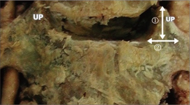
Fig. 1 Photograph of a cadaveric specimen showing the height and width of the uncinate process. 1 : height of uncinate process, 2 : width of uncinate process, UP : uncinate process.

Fig. 2 Photograph of the transverse section through the disc level of the cervical spine showing the width of the uncinate process and the distance between the medial borders of the uncinate processes from each side of the vertebra. 1 : width of the uncinate process, 2 : distance between medial borders of uncinate processes at anterior margin, 3 : distance between medial borders of uncinate processes at midpoint, 4 : distance between medial borders of uncinate processes at posterior margin, UP : uncinate process.

Fig. 3 Photograph of the transverse section through the disc level of the cervical spine. The relationship of the vertebral artery and nerve root to the uncinate process is shown. 1 : angle between posterior tip of uncinate process and vertebral artery, 2 : shortest distance from posterior tip of uncinate process to nerve root, 3 : shortest distance from lateral wall of uncinate process to vertebral artery, NR : nerve root, UP : uncinate process, VA : vertebral artery.
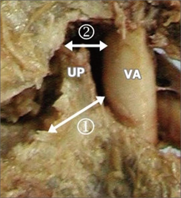
Fig. 4 Photograph of the cadaveric specimen showing the distance from the most anteromedial point of the uncinate process to the vertebral artery and the distance from the tip of uncinate process to the vertebral artery. 1 : distance from the most anteromedial point of uncinate process to vertebral artery, 2 : distance from upper tip of uncinate process to vertebral artery, UP : uncinate process, VA : vertebral artery.
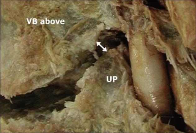
Fig. 5 Photograph of the cadaveric specimen showing Luschka joint space, corresponding to the distance from the upper tip of the uncinate process to the vertebral body immediately above it. UP : uncinate process, VB : vertebral body.
Reference
-
1. Bartolomei J, Sonntag V. Winn HR, Youmans JR, editors. Anterior approach including cervical corpectomy. Youmans' Neurological Surgery. 2004. ed 5. Philadelpia: WB Saunders;p. 4431–4445.2. Chang JC, Park HK, Bae HG, Cho SJ, Choi SK, Byun PJ. Morphometric measurement of the anatomical landmark in anterior cervical microforaminotomy. J Korean Neurosurg Soc. 2006; 39:340–346.3. Cooper PR. Anterior cervical vertebrectomy : tips and traps. Neurosurgery. 2001; 49:1129–1132. PMID: 11846907.4. Flynn TB. Neurologic complications of anterior cervical interbody fusion. Spine (Phila Pa 1976). 1982; 536–539. PMID: 7167824.
Article5. Hardy RW. Tarlov EC, editor. Complication of anterior cervical surgery. Complications of spinal surgery. 1991. Park Ridge: American Association of Neurological Surgeons;p. 85–94.6. Hirsch C. Cervical disc rupture : diagnosis and therapy. Acta Orthopaedica. 1961; 30:172–186.7. Horwitz NH, Rizzoli HV. Horwitz NH, Rizzoli HV, editors. Herniated intervertebral discs and spinal stenosis. Postoperative complications of extracranial neurosurgery. 1987. Baltimore: Williams & Wilkins;p. 30–98.8. Jang WY, Kim KS, Lee JC, Kim CJ, Choi HY, Xuan XN, et al. Results of microsurgical anterolateral tunnel approach for cervical disc herniation. J Korean Neurosurg Soc. 2001; 30:600–604.9. Jho HD. Microsurgical anterior cervical foraminotomy for radiculopathy : a new approach to cervical disc herniation. J Neurosurg. 1996; 84:155–160. PMID: 8592215.
Article10. Lu J, Ebraheim NA. The vertebral artery : surgical anatomy. Orthopedics. 1999; 22:1081–1085. PMID: 10580826.11. Luschka H. Die Halbgelenke des menschlichen korpers. 1858. Berlin: Reimer.12. Oh SH, Perin NI, Cooper PR. Quantitative three-dimensional anatomy of the subaxial cervical spine : implication for anterior spinal surgery. Neurosurgery. 1996; 38:1139–1144. PMID: 8727144.
Article13. Osborne AG. Diagnostic cerebral andiography. 1999. ed 2. Philadelphia: Lippincott Williams & Wilkins.14. Pait TG, Killefer JA, Arnautovic KI. Surgical anatomy of the anterior cervical spine : the disc space, vertebral artery, and associated bony structures. Neurosurgery. 1996; 39:769–776. PMID: 8880772.
Article15. Payne EE, Spillane JD. The cervical spine; an anatomico-pathological study of 70 specimens (using a special technique) with particular reference to the problem of cervical spondylosis. Brain. 1957; 80:571–596. PMID: 13499761.
Article16. Schneider RC. Schneider RC, Kahn EA, Crossby EC, Taren JA, editors. Treatment of cervical spine disease. Correlative neurosurgery. 1982. ed 3. Springfield: Charles C Thomas;p. 1094–1174.17. Schweighofer F, Passler JM, Wildburger R, Hofer HP. Interbody fusion of the lower cervical spine : a dangerous surgical method? Langenbecks Arch Chir. 1992; 377:295–299. PMID: 1405955.18. Shintani A, Zervas NT. Consequence of ligation of the vertebral artery. J Neurosurg. 1972; 36:447–450. PMID: 5013615.
Article19. Smith MD, Emery SE, Dudley A, Murray KJ, Leventhal M. Vertebral artery injury during anterior decompression of the cervical spine. A retrospective review of ten patients. J Bone Joint Surg Br. 1993; 75:410–415. PMID: 8496209.
Article20. Trolard P. [Quelques articulations de la colonne vertebrale]. Int Monatschr F Anat Physiol. 1893; 10:1–11.
- Full Text Links
-
- Actions
-
Cited
- CITED
-
- Close
- Share
-
- Similar articles
-
- Morphometry of the Uncinate Process, Vertebral Body, and Lamina of the C3–7 Vertebrae Relevant to Cervical Spine Surgery
- Anatomical variations and surgical implications of axillary artery branches: an anatomical study of the coracoid process region
- Unduly extensive uncinate process of pancreas in conjunction with pancreatico-duodenal fold
- Preoperative Radiographic Simulation for Partial Uncinate Process Resection during Anterior Cervical Discectomy and Fusion to Achieve Adequate Foraminal Decompression and Prevention of Vertebral Artery Injury
- Morphometrical Study of Uncinate Processes and Vertebral Body of Cervical Spine
