Sclerosing Epithelioid Fibrosarcoma of the Kidney
- Affiliations
-
- 1Department of Urology, Korea Cancer Center Hospital, Seoul, Korea. andrea@kcch.re.kr
- KMID: 2061303
- DOI: http://doi.org/10.4111/kju.2007.48.9.986
Abstract
- Sclerosing epithelioid fibrosarcoma(SEF) is an uncommon tumor of the deep soft tissue. It was first described in 1995 by Meis-Kindblom et al. Histologically, this tumor is characterized by uniform, small, round to ovoid epithelioid cells with clear cytoplasm, and the cells are arranged in distinct nests and cords. On immunohistochemistry, the most consistently positive marker is vimentin, although other antigens(cytokeratin, epithelial membrane antigen, S100 protein and neuron specific enolase) have been recorded as being inconsistently positive. We report here on a case of a 46-year-old woman who presented with back pain. The radiologic findings revealed a right renal mass and multiple bone metastases. The patient underwent radical nephrectomy and the pathologic finding was primary SEF of the kidney.
Keyword
MeSH Terms
Figure
-
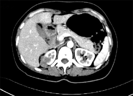
Fig. 1 Abdominal CT scan shows a 3cm sized lobulated enhanced solid mass in the upper pole of the kidney.
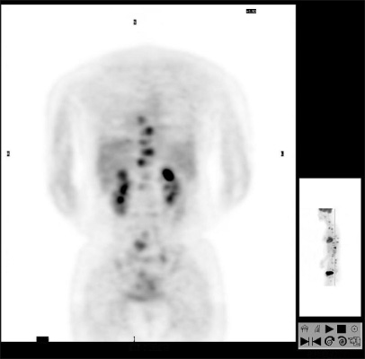
Fig. 2 Positron emission tomography (PET) shows hypermetabolic lesions in the right kidney (standardized uptake value=3.4), the CT-L-S spines, the ribs and the femur (standardized uptake value=4.1).
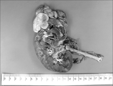
Fig. 3 Sagittal section reveals a well demarcated white to tan and firm mass that measured 4.0×3.0×3.2cm at the renal capsule.
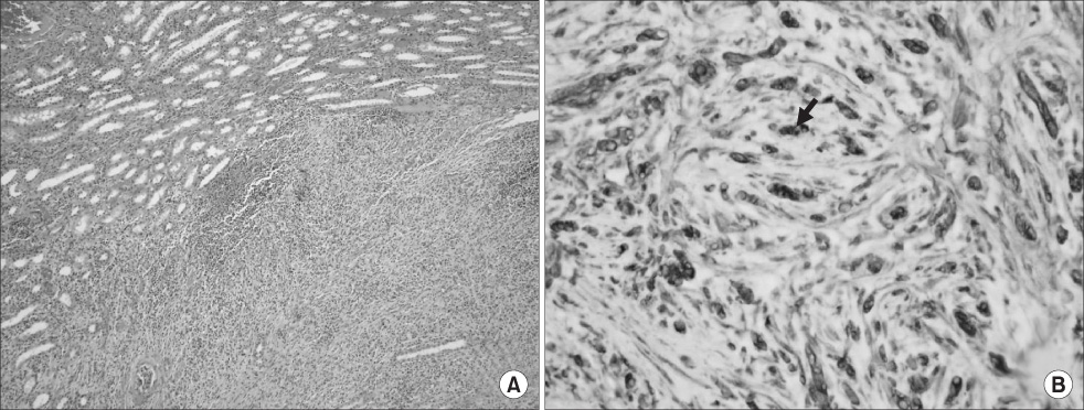
Fig. 4 Histologic and immunohistochemistry findings of sclerosing epithelioid fibrosarcoma (SEF). (A) The tumor cells are uniform, small, round to ovoid epithelioid cells with clear cytoplasm, and they are arranged in distinct cords and strands (H&E, ×100). (B) There is positive staining for vimentin. Arrow: positive staining (Vimentin, ×400).
Reference
-
1. Meis-Kindblom JM, Kindblom LG, Enzinger FM. Sclerosing epithelioid fibrosarcoma. A variant of fibrosarcoma simulating carcinoma. Am J Surg Pathol. 1995. 19:979–993.2. Eyden BP, Manson C, Banerjee SS, Roberts IS, Harris M. Sclerosing epithelioid fibrosarcoma: a study of five cases emphasizing diagnostic cirteria. Histopathology. 1998. 33:354–360.3. Antonescu CR, Rosenblum MK, Pereira P, Nascimento AG, Woodruff JM. Sclerosing epithelioid fibrosarcoma: a study of 16 cases and confirmation of a clinicopathologically distinct tumor. Am J Surg Pathol. 2001. 25:699–709.4. Watanabe K, Suzuki T. Epithelioid fibrosarcoma of the ovary. Virchows Arch. 2004. 445:410–413.5. Lee JG, Sohn KM, Kwon DD, Oh BR, Ryu SB, Park YI, et al. A case of fibrosarcoma of kidney. Korean J Urol. 2001. 42:770–772.6. Campbell SC, Novick AC, Bukowski RM. Kavoussi LR, Novick AC, Partin AW, Peters CA, Wein AJ, editors. Renal tumors. Campbell's urology. 2007. 9th ed. Philadelphia: Saunders;1567–1637.7. Bilsky MH, Schefler AC, Sandberg DI, Dunkel IJ, Rosenblum MK. Sclerosing epithelioid fibrosarcomas involving the neuraxis: report of three cases. Neurosurgery. 2000. 47:956–959.8. Abdulkader I, Cameselle-Teijeiro J, Fraga M, Caparrini A, Forteza J. Sclerosing epithelioid fibrosarcoma primary of the bone. Int J Surg Pathol. 2002. 10:227–230.
