Parathyroid Cyst Presenting as Acute Pancreatitis: Report of a Case
- Affiliations
-
- 1Department of Internal Medicine, Chonnam National University Medical School, Gwangju, Korea. yejoo@chonnam.ac.kr
- 2Department of Pathology, Chonnam National University Medical School, Gwangju, Korea.
- KMID: 2048817
- DOI: http://doi.org/10.4068/cmj.2013.49.3.125
Abstract
- We report the first case of hypercalcemia-induced acute pancreatitis caused by a functioning parathyroid cyst in a 67-year-old man. Laboratory investigation revealed increased serum amylase and lipase, increased serum ionized calcium and parathyroid hormone (PTH) levels, and decreased serum phosphate, indicating pancreatitis and primary hyperparathyroidism (PHPT). Abdominal computed tomography (CT) revealed mild swelling of the pancreatic head with peri-pancreatic fat infiltration and fluid collection around the pancreatic tail. Ultrasonography and CT of the neck showed a cystic lesion at the inferior portion of the left thyroid gland, suggesting a parathyroid cyst. There was no evidence of parathyroid adenoma by 99mTc sestamibi scintigraphy. PHPT caused by a functioning parathyroid cyst was suspected. The patient underwent surgical resection of the functioning parathyroid cyst owing to his prolonged hypercalcemia. At 3 weeks after the operation, his serum levels of PTH, total calcium, ionized calcium, inorganic phosphate, amylase, and lipase were normalized. At the follow-up examinations, he has remained asymptomatic.
Keyword
MeSH Terms
Figure
-
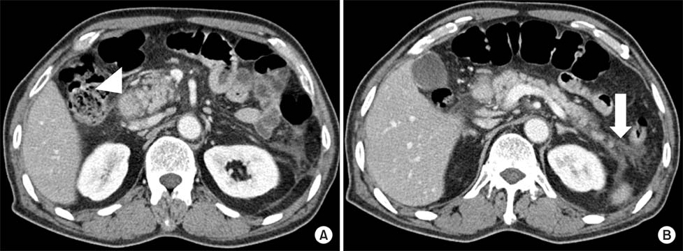
FIG. 1 Abdominal computed tomography (A, B) showed mild swelling of the pancreatic head with peri-pancreatic fat infiltration (arrow head) and fluid collection around the pancreatic tail (arrow).
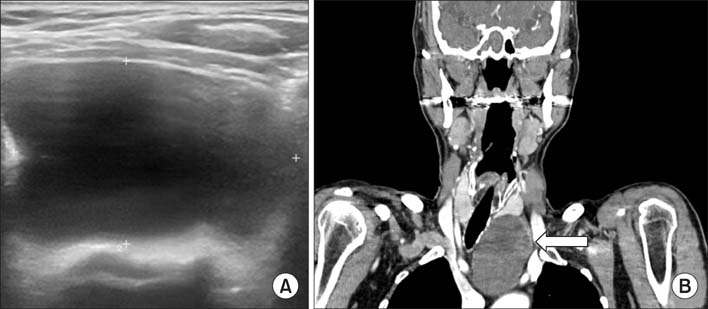
FIG. 2 Neck ultrasonography revealed a left infrathyroidal cystic lesion (A). Neck computed tomography revealed an approximately 5.4-cm sized low attenuated lesion (arrow) in the left the infrathyroidal area extending to the mediastinum (B).
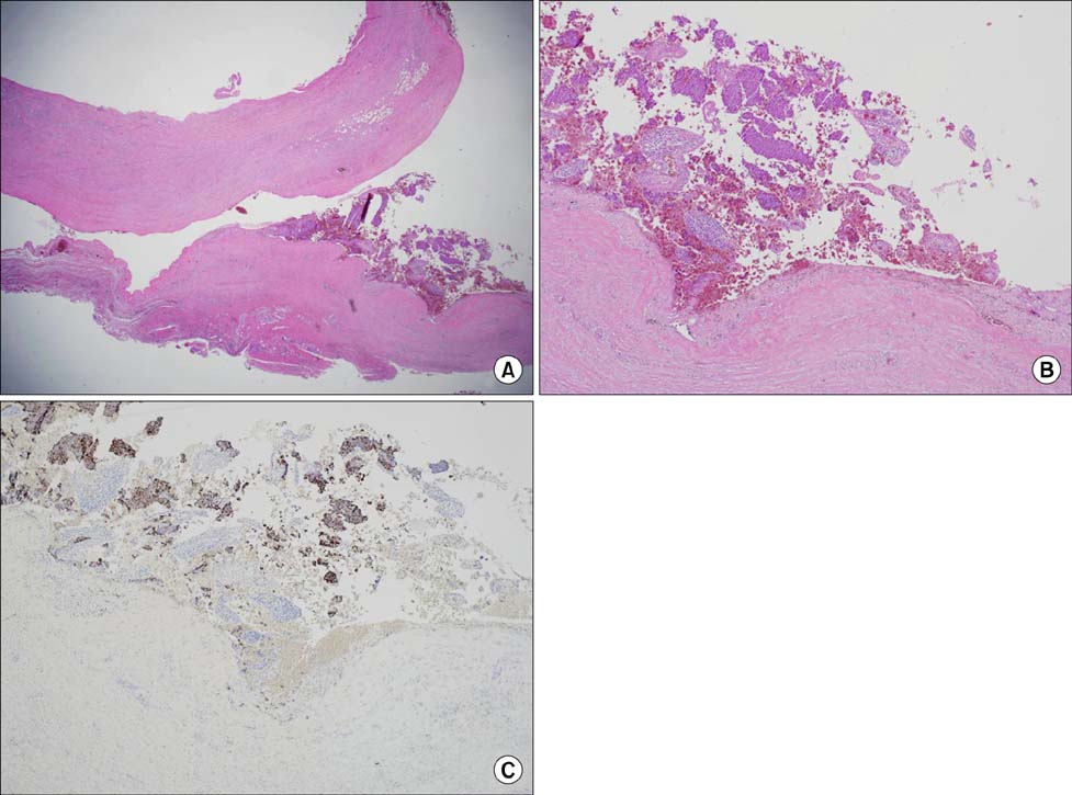
FIG. 3 Histopathological examination of the cyst revealed no epithelial lining and a cystic wall composed of fibrous tissue (hematoxylin&eosin stain, ×12.5) (A), (H&E stain, ×40) (B). Focal parathyroid hyperplasia along the luminal side of the cyst wall was positive for parathyroid hormone (PTH stain, ×40) (C).
Reference
-
1. Whitcomb DC. Clinical practice. Acute pancreatitis. N Engl J Med. 2006; 354:2142–2150.2. Bai HX, Giefer M, Patel M, Orabi AI, Husain SZ. The association of primary hyperparathyroidism with pancreatitis. J Clin Gastroenterol. 2012; 46:656–661.
Article3. Egea Valenzuela J, Belchí Segura E, Sánchez Torres A, Carballo Alvarez F. Acute pancreatitis associated with hypercalcemia. A report of two cases. Rev Esp Enferm Dig. 2009; 101:65–69.
Article4. Fraser WD. Hyperparathyroidism. Lancet. 2009; 374:145–158.
Article5. Gough IR. Parathyroid cysts. Aust N Z J Surg. 1999; 69:404–406.
Article6. Frick TW. The role of calcium in acute pancreatitis. Surgery. 2012; 152:3 Suppl 1. S157–S163.
Article7. Imachi H, Murao K, Kontani K, Yokomise H, Miyai Y, Yamamoto Y, et al. Ectopic mediastinal parathyroid adenoma: a cause of acute pancreatitis. Endocrine. 2009; 36:194–197.
Article8. Delaunay T, Peillon C, Manouvrier JL, Deotto JF, Doucet J, Nicaise JM, et al. Cysts of the parathyroid glands. Apropos of 6 cases. Ann Chir. 1990; 44:231–235.9. Rosenberg J, Orlando R 3rd, Ludwig M, Pyrtek LJ. Parathyroid cysts. Am J Surg. 1982; 143:473–480.
Article10. Khan A, Khan Y, Raza S, Akbar G, Khan M, Diwan N, et al. Functional parathyroid cyst: a rare cause of malignant hypercalcemia with primary hyperparathyroidism-a case report and review of the literature. Case Rep Med. 2012; 2012:851941.
Article
- Full Text Links
-
- Actions
-
Cited
- CITED
-
- Close
- Share
-
- Similar articles
-
- Pancreatitis-Primary Hyperparathyroidism Association: Case Report and Literature Review
- A Case of Parathyroid Adenoma Manifested by Acute Recurrent Pancreatitis
- A Case of Mediastinal Cystic Parathyroid Adenoma Presenting as Acute Pancreatitis
- A Case of Pancreatitis Associated with Hyperfunctioning Intrathyroidal Parathyroid Adenoma
- A Case of Parathyroid Adenoma Presenting as Acute Pancreatitis Accompanied with Empty Sella
